


1. Introduction
Polyhexamethylene guanidine (PHMG) is a positive charged oligomeric and polymeric compound that has been applied for many years in several industries [1, 2]. It contains multiple guanidine groups and is known as a potent biocide with a broad spectrum of bioactivities [3, 4]. When PHMG was produced as a household product, its toxicity to the human body was underestimated due to poor or wrong evaluation experiments, making many people in Korea suffer from a tragic outbreak of severe pulmonary disorders. The lung diseases originated from the use of PHMG as a humidifier disinfectant [5–8]. For more than 10 years, approximately 200 people have died, among them children and pregnant women as well, from the inhalational toxicity of PHMG. Moreover, according to the press release reported by the Ministry of Environment, several hundred people continue to have serious respiratory diseases caused by the humidifier disinfectant [9]. A lot of studies have confirmed that the inhalation of PHMG caused lung injuries, such as acute inflammation and catastrophic fibrogenesis [10–13]. In addition, various studies have been conducted or are under investigation to verify the toxicity of PHMG to other internal organs of the human body [14–18].
To evaluate organ toxicity on exposure to toxic substances, its uptake, distribution, and excretion profile need to be determined in a living subject [19, 20]. However, the measurement of the biological uptake of PHMG is difficult due to the limited detection techniques in tissues, which contain complex chemical species and biomolecules. Mass spectrometry cannot be used to analyze PHMG in biological samples since PHMG comprises a mixture of polymers with many molecular weights. In addition, this material does not generate a convenient optical signal nor adsorption spectrum, and thus ultraviolet–visible and fluorescent detectors would not be useful to detect trace quantities in vivo. These limitations can be overcome using a radionuclide as a tracer. The radiation energy (i.e., γ-ray) emitted from a conventionally used radionuclide in nuclear medicine can penetrate biological tissues without any signal interference. Therefore, the method employing a radiotracer allows straightforward, accurate, and sensitive detection, as well as non-invasive imaging in a living subject [21–25]. In the previous study, we have reported the radiolabeling of PHMG to determine uptake values in animal models [26]. The radioactive indium (111In)-labeled PHMG aerosol was exposed to Sprague–Dawley (SD) rats by inhalation, following which the toxic chemical in biological tissues was measured. The high accumulation and retention of PHMG aerosols in the lungs, as well as transport from the lungs, were observed quantitatively through the experiment. Although these results suggest the severe inhalation toxicity of PHMG, further in vivo assessment is still necessary for an in-depth understanding of its effect in other internal organs. In this study, we report the biodistribution of radiolabeled PHMG following intravenous injection for 1 week to investigate organ-specific uptake, retention, and clearance of PHMG. Then, the results will be discussed by comparing other organ distribution results obtained by the intratracheal instillation and oral administration, which are considered main exposure routes of PHMG. In addition, non-invasive whole-body images are provided to visualize PHMG administered in animal models. Fig. 1 illustrates the study design, which includes the organ distribution study and single-photon emission computed tomography (SPECT) imaging using the radiolabeled PHMG.
2. Materials and Methods
2.1. General Procedure
The radioisotope ([111In]InCl3) was supplied by Mallinckrodt Pharmaceuticals (Netherlands). The PHMG solution (25 wt% in water, SKYBIO 1125, weight average molecular weight = ca. 4.1 kDa) was provided by the Korea Institute of Toxicology (Jeongeup, Republic of Korea). All commercially available reagents were used without further purification. For the preparative high-performance liquid chromatography (HPLC) experiment, 0.1% formic acid containing H2O (solvent A), and 0.1% formic acid, containing acetonitrile (solvent B), were used as eluents (Agilent 1260 Infinity, USA). The flow rate was 10 mL/min for preparative HPLC (column: Eclipse XDB-C18, 21.2 × 150 mm, 7 mm, eluent gradient: 100% solvent A for 0–5 min; 30% solvent B in solvent A for 5–20 min; 70% solvent B in solvent A for 20–25 min; 100% solvent A for 25–26 min; retention time of PHMG–DOTA: 20.0–24.5 min). The amount of radioactivity was measured using γ-counter (1480 Wizard 3, PerkinElmer, USA). The radiolabeling reaction was monitored using a radio-thin-layer chromatography (radio-TLC) imaging scanner (AR-2000, Eckert & Ziegler, USA). The SPECT images were obtained using a small animal SPECT/PET/CT system (Siemens, USA).
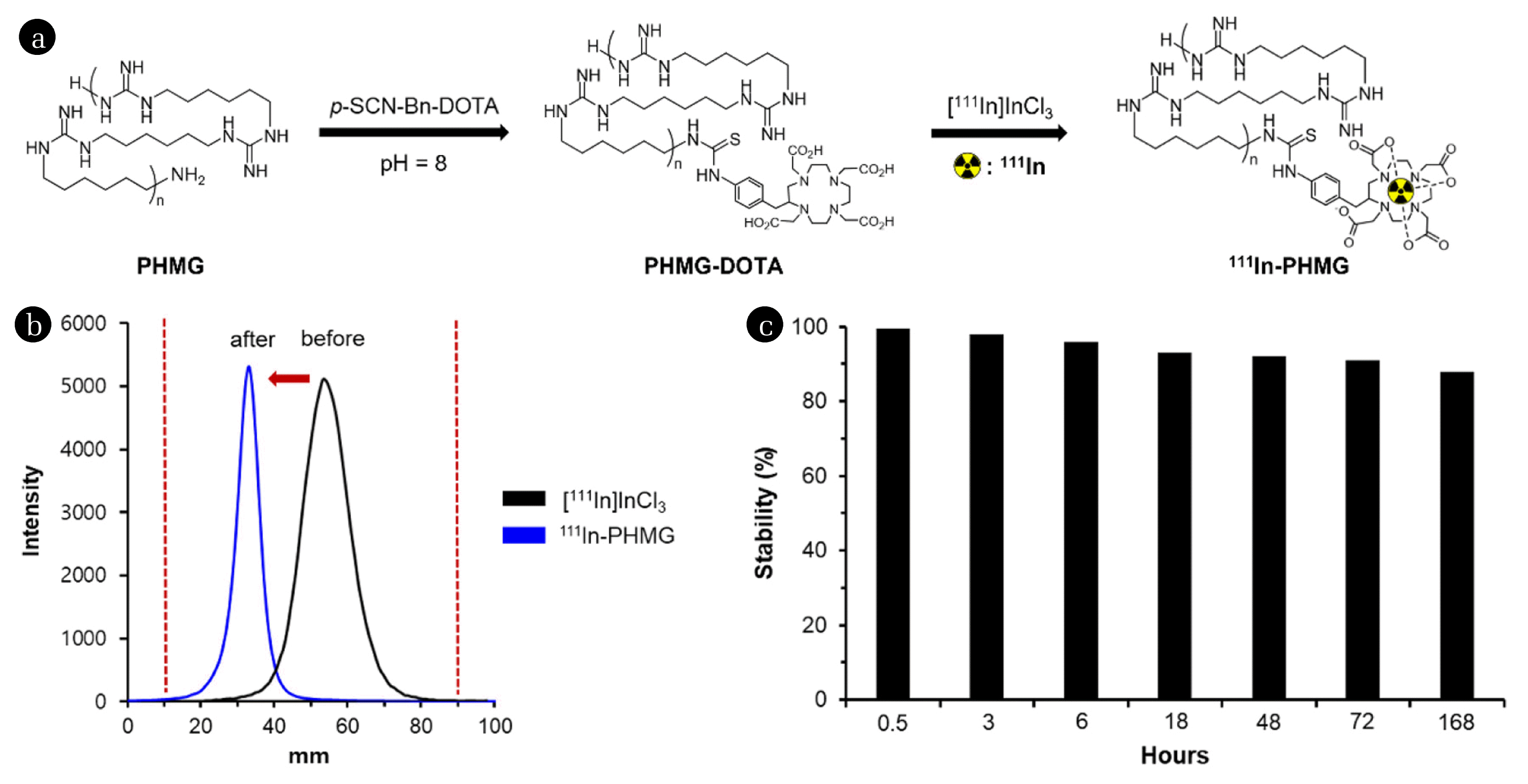
2.2. Preparation of Polyhexamethylene Guanidine (PHMG)–DOTA and Radiolabeling of PHMG
The conjugation of PHMG with DOTA (1,4,7,10-tetraazacyclodecane-1,4,7,10-tetraacetic acid) and 111In labeling was conducted following the methods in our previous report [26]. Aqueous PHMG (25 wt%, SKYBIO 1125) reacted with three equivalent of p-SCN-Bn-DOTA for 6 h at 37°C (pH = 8). Unreacted or hydrolyzed DOTA in the crude mixture was removed using preparative HPLC purification. The product was characterized using nuclear magnetic resonance spectroscopy. PHMG–DOTA conjugate was radiolabeled by adding 148 MBq of [111In]InCl3 in 0.1 M aqueous HCl to 1 mg of PHMG–DOTA in 50 μL pure water. The pH was adjusted to 4 using aqueous sodium hydroxide, and the resulting mixture was heated at 80°C for 15 min and then cooled to 25°C. Radiolabeling was monitored using radio-TLC, as shown in Fig. 2(b). The radiochemical purity of the final product (111In-PHMG) was determined using radio-TLC.
2.3. Animal Care for in Vivo Experiments
Male SD rats (aged 8 weeks, 250–270 g) obtained from Orient Bio Inc. (Iksan, Republic of Korea) were used for in vivo imaging and biodistribution experiments. Animals were randomly divided into seven groups (five rats per group) for biodistribution studies. They were kept in ventilated cages under standard conditions of temperature, humidity, pressure, and light (light/dark cycle for 12 h). Animals were provided with a sterile diet and water ad libitum. The protocols of animal studies were approved by the Institutional Animal Ethical Committee (Korea Atomic Energy Research Institute) and performed under strict compliance with the guidelines issued by the committee.
2.4. Biodistribution Study of 111In-labeled Polyhexamethylene Guanidine
111In-PHMG (37 KBq/100 μL) in saline was administered to SD rat body (weight: 250–270 g) through different administration routes (intravenous injection, intratracheal instillation, oral administration). At each time point (0.5, 3, 6, 18, 48, 72, and 168 h), five rats were sacrificed under anesthesia and the organs (heart, lungs, liver, spleen, kidneys, stomach, small intestine, large intestine, and thyroid) and the blood were harvested. The radioactivity of the collected organs was measured using γ-counter. The distribution data were shown as percentages of injected dose (%ID) or percent of injected dose per gram of tissue (%ID/g).
2.5. Single-photon Emission Computed Tomography/Computed Tomography Imaging of 111In-labeled Polyhexamethylene Guanidine
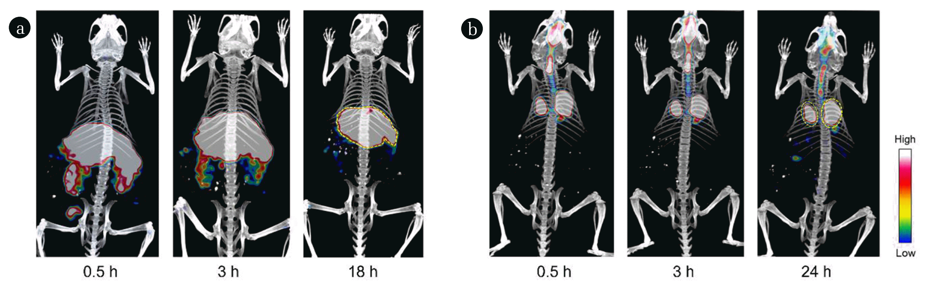
The animal was anesthetized with 2% isoflurane. 111In-PHMG (5.6 MBq/100 μL) solution was administered through different administration routes (intravenous injection and intratracheal installation). SPECT/computed tomography (CT) images were obtained using a small animal SPECT/CT system (Siemens, USA) at 0.5, 1.5, and 18 h (or 24 h) post administration.
3. Results and Discussion
3.1. Preparation of Radiolabeled Polyhexamethylene Guanidine (PHMG) (111In-PHMG)
To introduce 111In to the PHMG molecule, the amino group of PHMG was first conjugated with DOTA (Fig. 2(a)). For efficient incorporation of a metal chelating agent, three equivalents of DOTA-isothiocyanate were mixed with PHMG solution for 6 h at pH = 8 and 37°C. At the end of the reaction, the crude product was purified, using a preparative HPLC, to give DOTA conjugated PHMG (PHMG-DOTA). The radiolabeling was performed by mixing an aqueous solution of PHMG-DOTA with [111In]InCl3 for 15 min at pH = 4 and 80°C. Radio-TLC analysis showed that the Rf value of free 111In metal was 0.55, whereas that of radiolabeled PHMG (111In-PHMG) was 0.29 when aqueous citric acid (0.1 M) was used as an eluent (Fig. 2(b)). The radiolabeling was accomplished with a high radiochemical yield and the amount of unbounded radioactive indium ([111In]InCl3) in the reaction mixture was negligible. As the radiolabeling procedure was straightforward and provided high radiochemical purity (> 99%), analyzed by radio-TLC, no further purification was necessary for the next experiments.
3.2. In vitro Stability Studies
The stability of the radiolabeled PHMG was tested in mouse serum at 37°C. In this study, 10 μL (3.7 MBq) of the desired 111In-PHMG was incubated with 90 μL serum in a microcentrifuge tube (0.6 mL) at the abovementioned temperature. The amount of intact 111In-PHMG was determined using radio-TLC at 0.5, 3, 6, 18, 48, 72, and 168 h. However, the results of the radio-TLC chromatogram were confusing, as the metabolized or protein-bounded radiotracer showed broad Rf values, which impede accurate integration for analyzing the stability in the serum. Thus, radio-HPLC was used to determine the serum stability of 111In-PHMG. The retention time of free 111In metal was less than 3 min, which makes a large difference in the retention time than the 111In-PHMG. The liberated free 111In after 168 h incubation in the serum was as low as 12% and approximately 88% of 111In was retained with the parental molecule (Fig. 2(c)), which is enough stability for in vivo study.
3.3. Biodistribution Studies in Sprague–Dawley Rats
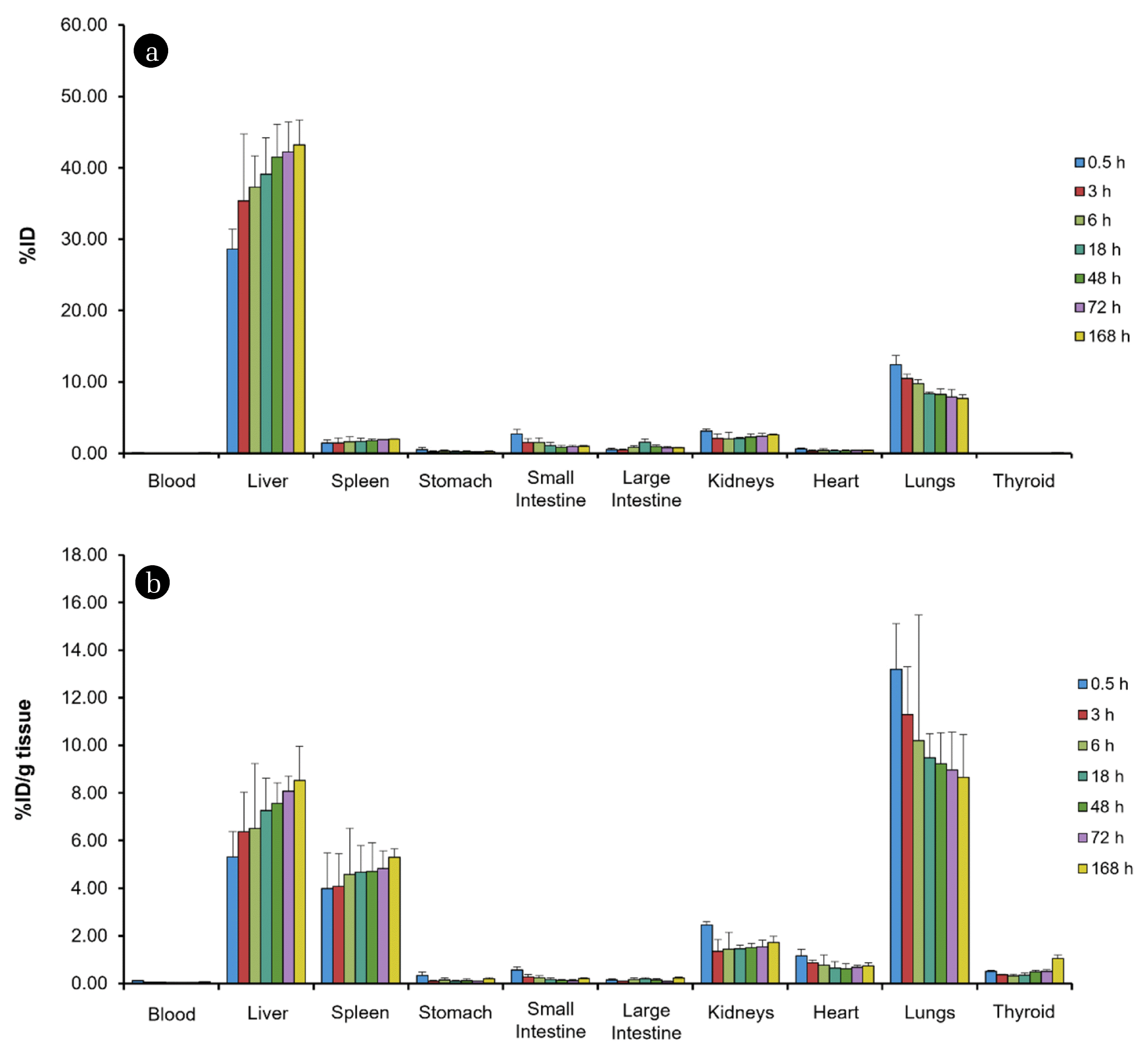
The biodistribution studies of 111In-PHMG were conducted in male SD rats. The radiolabeled PHMG was diluted in saline (aqueous 0.9% NaCl solution) to a final concentration of 37 KBq/100 μL. The radiotracer was then administered to animals through three routes (intravenous injection, intratracheal instillation, and oral administration). The blood and nine organs were dissected and analyzed seven times, up to 168 h post-exposure, using γ-counter for quantitative measurement of PHMG in biological tissues. The radioactivity in excreta (urine and feces) was not collected in this experiment; thus, the biodistribution data would not represent all the activity administered.
The amount of PHMG distributed in tissues and blood is shown in Fig. 3 after decay correction of radioactivity at the end of intravenous injection (t = 0 h). The biodistribution data plotting the %ID (Fig. 3(a)) exhibited the highest uptake in the liver (28.63%ID at 0.5 h post-exposure) and the levels were increased over time. A similar trend was observed in the spleen, which represents the accumulation of PHMG in these organs. PHMG is highly distributed in the lungs (12.44%ID at 0.5 h post-exposure), and the observed radioactivity decreased slowly within 168 h. The uptake values in the lungs were to be more remarkable when the radioactivity was normalized to tissue mass (%ID/g, Fig. 3(b)). At the initial time point, a noticeable uptake of 111In-PHMG was found in the lungs with 13.19%ID/g, and this amount was higher than those of the liver (5.31%ID/g) and spleen (3.99%ID/g). In addition, only approximately 35% of the initial uptake value (0.5 h) in the lungs was reduced after 168 h post-injection (8.65%ID/g). However, the radioactivity observed in the small and large intestines, and kidneys, which include excretory organs, were lower than in the liver and lungs. These results suggest high retention in the internal organs, as well as extremely slow clearance kinetics of intravenously injected PHMG.
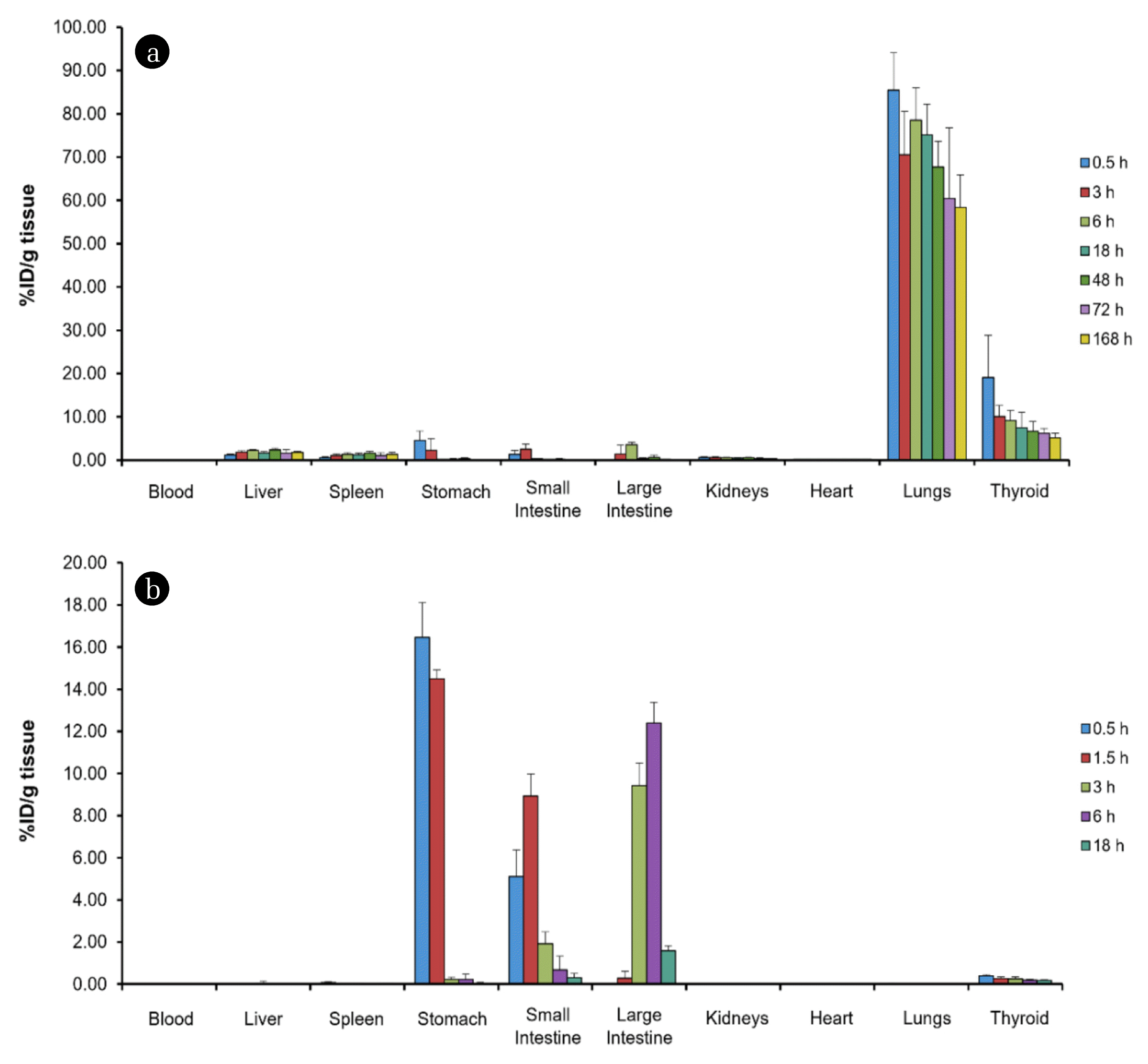
In addition, we investigated the biodistribution after 111In-PHMG was exposed to SD rats through intratracheal instillation to observe the retention of PHMG in the lungs and the amount of translocation from the lungs. Radioactivity was mostly observed in the lungs at the initial time point (0.5 h, 85.47%ID/g). The data demonstrated that administered PHMG was cleared slowly, with approximately 68% of radioactivity remaining in the lungs after 168 h. Some PHMG was translocated to the extrapulmonary organs by passing the radiotracer through the air–blood barrier in the respiratory system [27, 28]. The quantification of radioactivity in some tissues exhibited PHMG retention over time. For example, the uptake values of the liver were 1.12 and 1.74%ID/g at 0.5 and 168 h post exposure, respectively. In addition, an increase in uptake values was detected in the spleen. These trends were similar to those observed in Fig. 3, which exhibited persistent retention of PHMG in the lungs and liver, following intravenous administration. Taken together, prolonged exposure of PHMG through the respiratory system would have fatal effects to the lungs as well as other internal organs. The humidifier disinfectant can also be exposed to human via oral route. Interestingly, orally administered PHMG was cleared out in a day without significant translocation to other internal organs (i.e., lungs, heart, liver, and spleen) (Fig. 4(b)) from intestines. This result indicated that PHMG observed in gastrointestinal organs would have little effect on the uptake in the extraintestinal organs including lungs. The uptake values in each organ and the clearance rate could be related to the toxicity of PHMG. Our results in the present study are consistent with the recent toxicological studies, reporting that the oral lethal dose (LD50) of PHMG for rats is 610 mg/kg of body weight [29]. However, inhalation exposure is much more fatal, because the inhalation LD50 is observed to be 0.094–0.155 mg/L when it is exposed to rats for 4 h.
Next, the biological distribution of PHMG was investigated through the molecular imaging method. Whole-animal SPECT images were obtained for 24 h post administration. For 111In-PHMG injected intravenously (Fig. 5(a)), a strong signal was detected in the internal organs, whereas the radiotracer exposed to the respiratory tract showed localized accumulation in the lungs and the intensity of the signal was retained for 1 day (Fig. 5(b)), which is consistent with the biodistribution results. In contrast, the residual 111In-PHMG exposed using oral administration was restricted to the gastrointestinal tract, and the signal in the whole body decreased after 24 h exposure to the radiotracer [26].
Recent studies reveal the exposure of PHMG induced dose- and time-dependent cytotoxicity in various cells [9, 30–32]. The cationic guanidine groups in PHMG could bind negatively charged cellular components and biomolecules (e.g., phospholipids and proteins), which disrupted the membrane integrity, blocked the enzymatic pathway, and caused toxic effects on cell metabolism and survival [33, 34]. In addition, PHMG could consider interacting with intracellular organelles, which produce reactive oxygen species and lead to apoptosis [35]. According to the results in the present experiments, a part of intratracheally administered PHMG can be translocated to extrapulmonary organs. Moreover, the amount of PHMG distributed in some internal organs, such as the liver, spleen, and kidneys, did not significantly decrease for 1 week, which showed an unusual execration pattern. Considering that a single administration of radiolabeled PHMG in this study showed considerable accumulation of toxic chemicals, repeated exposure of PHMG should be responsible for severe toxicity in the respiratory system, as well as internal organs. The observed uptake values and SPECT imaging results would provide useful information to understand the toxicological effects of PHMG in various organs.
4. Conclusions
In this study, we demonstrate the radiolabeling of PHMG using 111In to measure its biological uptake in animal models. The radiotracer showed acceptable stability under the physiological conditions, and thus it was used to investigate the biological distribution of PHMG exposed to three administration routes. The results revealed that intravenously injected PHMG accumulated to a high level in the internal organs, suggesting that continual exposure to PHMG can be highly toxic to the human body. In addition, the data evidenced the transport of inhaled PHMG, which may be used to investigate the toxicological effects on extrapulmonary organs. The radio-analytical methods and molecular imaging strategy in this study can be used to investigate the risk assessment of other toxic polymers and disinfectants. Moreover, our approach can be used to assess the bioaccumulation and in vivo behavior of emerging contaminants, including inhaled fine particulate matter and ingested microplastics.