AbstractIn the present study we have prepared graphene supported CeO2-TiO2 mixed oxide and studied its activity towards photocatalytic degradation of methylene blue (MB) dye. The mixed oxide is prepared with the variation of cerium loading to that of titanium from 2% to 10% by sol-gel method and graphene oxide (GO) is prepared by using modified Hummer’s method. The resulting adsorbent nanocomposite is synthesised by chemical reduction method with GO loading 2% by weight to that of mixed metal oxide and employed in the catalytic process through batch wise adsorption to test the optimum level of activity by varying the parameters (pH: 3–10, time: 15–75 min, dye concentration: 5–40 ppm, anions like Cl−, SO42−, NO3−, HCO3−) with catalyst loading 0.020 g per 0.100 L of the dye solution. It is inferred that the adsorbent catalyst nanocomposite with ceria loading up to 5% with that of titania has better catalytic activity towards the photodegradation of MB with the optimum condition of pH-10 and time-75 minutes. Degraded product characterisation is carried out by LC-MS technique, which confirmed the degradation of MB. The adsorption kinetics is being investigated through pseudo-first-order kinetics and Langmuir adsorption model with good fitting having R2 value 0.986 and 0.993 respectively.
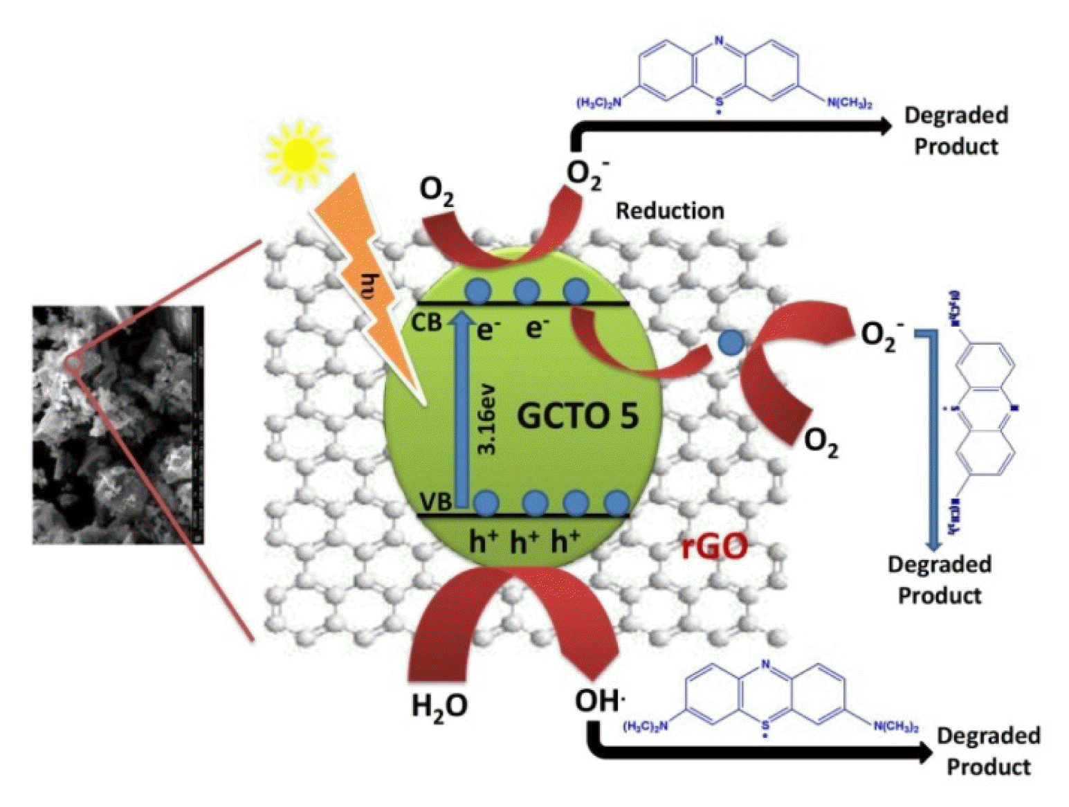
Graphical Abstract
1. IntroductionTopics related to energy and environment is one of the core research interests. The growing man-made industry has created an adverse effect in the habitat we live today. Over the decades, environmental pollution is a hot and demanding issue in the field of environmental remediation and pollution control. Wastewater generated from industries loaded with toxic organic compounds is one of the potent environmental pollutants. The disposal of harmful pollutant is a matter of concern for the environment. The main sources of environmental pollution are poor management of waste products including toxic chemicals [1–4]. Without proper treatment of these toxic loaded pollutants, waste disposal directly to the environment is matter of concern.
Industrial wastewater discharges have a relentless effect on the habitat we live in and adversely affect the health condition also. Organic dyes produced from textile industry are found to be one of chief pollutants among the industrial wastewaters. These dyes are highly toxic having a potent to cause cancer and due to their mutagenic essences, the food chain is interrupted leading to bioaccumulation. These dyes are soluble in water and drain the oxygen content affecting the penetration of light hence the aquatic life is adversely affected by disturbing the ecology. Cationic dyes including methylene blue utilized for dying mainly have adverse health effects like mental disorder, cardiac arrest, jaundice, nauseous etc. Complex aromatic structure with its synthetic genesis has displayed high stability under light and biodegradation. So, the usual water treatment process cannot be utilized for the removal of these dyes from the wastewater [5, 6]. For this, there is a grave demand to develop and study various methods for the treatment of industrially discharged wastewater constituting organic dyes. Several methods including oxidation [7], reduction, and flocculation [8] have been taken up for removing pollutants from wastewater. Prior to other methods employed photocatalysis stands out with better result. A number of semiconductors can also be used to degrade a variety of organic pollutants as depicted in Table 1 [9–30] using irradiation of light with different wave lengths. Among these technique photocatalysis has its prominent and challenging era in this category of environmental application [31, 32]. Semiconductor based photocatalysis is commercially available as it is cost effective and eco-friendly for discharging the wastewater. In photo catalysis there is no call for secondary disposal which stands out for water decontamination [33].
Among the metallic oxide, TiO2 is successfully employed in many industries for photocatalysis phenomenon because of its excellent photocatalytic activities, with economical availability and also due to the good chemical stability. This metallic oxide also shows an excellent restoration to the environment [34, 35]. Nevertheless, there are some limitations for employing TiO2 as photocatalyst. Having a wide band gap is the main de-merit in the photocatalysis as this is functional only in the UV region. The band energy gap is more than 3.2 eV with narrow excitation wavelength affecting the high rate of recombination of photo induced electron hole pair (e−& h+) and thus showing poor adsorption capacity. With such a wide band gap ranging from 3.2 to 3.5 eV [36, 37] some material gets activated by UV light which ultimately affecting the photocatalytic activity.
In order to reduce this band gap and lowering the light to the electron hole in TiO2 the composite metals are doped with several other metals like copper, gold, silver, nickel etc. non-metals including graphene, carbon nanotubes and several semiconductors are made available for the photocatalysis [38]. For this, several mixed oxides have been synthesised and the photocatalytic effects are considered [39].
Amid the rare elements, ceria displays consistent activities in the visible region of light when mixed with TiO2. Mostly cerium is present in nature in ample amount. Redox and catalytic activities with oxygen defects, its oxide form is employed in fuel cells as well for the pollution control analysis. The catalytic properties of CeO2 are due to the formation of Ce3+ defect sites with oxygen vacancies. CeO2 is a semiconductor with n-type and the band gap ranging from 2.7 to 3.4 eV [40]. Ceria-titania acts as an excellent photo catalyst due to several reason as offering thermal stability, lager surface area, economically and environmentally friendly. The important benefit of this material is its absorbance in the visible region of light [41, 42]. But the issue arises in poor conductivity, low energy band gap and blocking of the site with recombination of electron hole [43] which affects the photocatalysis. Some literatures have reported the formation of inter-band states when Ce is doped into the lattices of TiO2 during the exposure to visible light [44, 45]. As the size difference of Ce (in between 0.093 nm to 0.103 nm) to that of Ti (0.068 nm) is large, Ce cannot be fitted inside in the lattices of TiO2. In order to suppress these demerits several studies are carried out.
In the recent time to enhance the activities of photocatalytic nature of TiO2 by visible light, the modification of TiO2 is performed with carbonaceous substances such as carbon nanotubes, graphene to form carbon-TiO2 composites. To be particular in these carbonaceous substances, graphene stands out with good results as a material for photocatalysis due to its large surface area, flexible structure with high chemical stability and electrical conductivity. In order to enhance the photocatalytic activity, graphene-TiO2 composites are formed with aspiration of light absorption of TiO2 from UV region and thus increasing the photocatalytic activity of the compound resulting further degradation. The attractive feature of the graphene is due to its high energy storage and photocatalysis. Some of the properties such as high surface area with mechanical flexibility and thermal conductivity grasp graphene as the effective material for photocatalysis phenomenon. For this graphene finds application in photonic device as well as in optoelectronics. Graphene also has great contribution in the electromechanical properties of the device [46]. Hence graphene is a preferable material for improvement of the hetero junction-based photo detector in broad wavelength region.
The organic pollutant, methylene blue (MB) being its unique property of solubility in wide varieties of solvent like methanol, 2-propanol, water, ethanol, acetone, and ethyl acetate put our attention to carryout photocatalytic degradation study in our work. It is one type of cationic dye with molecular weight 319.85 g/mol and having absorption maxima at about 664 nm (parent peak) 612 nm (shoulder peak) and forming a stable solution with water at room temperature. The π-electron cloud present with conjugation in the nucleus is susceptible towards oxidation-reduction forming blue-colourless transformation respectively. It has wide range of application in textile, paper, dyeing and paint industries and used as a popular colourant material.
In this present work, we have reported the synthesis of graphene supported ceria-titania mixed oxide by sol-gel method and evaluated the photo catalytic activity towards MB degradation. The novelty of synthesis and photo catalytic activity of the nano sized material is explained with the help of various spectroscopic techniques and characterisation procedures.
2. Experimental2.1. Materials and MethodsAll the chemicals and reagents used are of analytical grade and used without further purification. Ceric ammonium nitrate (NH4)2Ce(NO3)6, titanium isopropoxide Ti(O-i-C3H7)4, graphite powder, propanol (C3H7OH), sodium nitrate (NaNO3), potassium permanganate powder (KMnO4), sulphuric acid (H2SO4), hydrogen peroxide (H2O2), deionised water (DIW) are used for the sample preparation.
2.2. Preparation of Mixed OxideThe different metal oxides are prepared by varying the weight ratio of cerium to that of titanium by stoichiometrically modifying one of our previous group work [47] and the work by Alberoni C et al. [48] to get the resulting metal oxide composites as CexTi(1−x)O2 where X=0.02, 0.05, 0.10 and labelled and depicted as CTO 2, CTO 5, CTO 10 respectively. In a typical method CTO 2 is prepared by dissolving a known quantity of (NH4)2Ce(NO3)6 in 25 ml deionised water. Stoichiometric quantity of C12H28O7Ti (titanium isopropoxide) is diluted to 30 ml by adding propanol. The ceric ammonium nitrate and titanium isopropoxide are added drop by drop to 450 ml water by maintaining pH 2.5 with constant stirring for 4 hours. Then the filtrate is washed with distilled water for several times and dried at 100°C. The sample is calcined at 400°C for 4 hours.
2.3. Preparation of Graphene OxideThe preparation of graphene oxide (GO) is carried out by using modified Hummers method. For graphene oxide preparation 1 g of graphite powder is mixed with 46 ml of conc. H2SO4 with stirring for 15 minutes followed by addition of 1 g NaNO3 and continued stirring for 30 minutes. Afterwards by adding 8 g of KMnO4, the solution is stirred for 2 hours at 20°C. Then 80 ml of DIW is added slowly to the composition and it is magnetically stirred continuously for 30 minutes. Then 30% H2O2 is added drop by drop till the effervescence is ceased. Then the mixture is washed with dilute HCl and DIW for several times. Finally, it is subjected to ethanol wash and water wash with centrifugation and the final product is dried at 80°C for 24 hours to confirm the identity of GO.
2.4. Preparation of Mixed Oxide CompositesThe mixed oxide composite is prepared by graphene to mixed metal oxide ratio with 2% GO by weight along with adopting chemical reduction method. The composite is prepared by dispersing calculated amount of GO in DIW and ultra-sonicated for 30minutes. To this dispersion, a known quantity of CTO 2 is added to keep the graphene loading in the final sample at 2% by weight and continued sonication for 30 minutes. After this, the suspension is stirred over a magnetic stirrer and sodium borohydride solution is added drop wise till the reaction stopped in order to have the reduced graphene oxide (r-GO) based metal mixed composite. Finally, it is centrifuged, dried and kept for further use. The sample is designated as GCTO 2. In this manner other samples GCTO 5 and GCTO 10 are prepared, and study is being carried out with graphene loading to only 2% by weight to avoid agglomeration or bending of graphene structure.
2.5. Preparation of Methylene Blue Stock SolutionActual weighed quantity 1 g of methylene blue (MB) (SRL India-Analytical grade) is dissolved in 100 ml of DIW and it is further diluted with DIW by adjusting the volume to 1000 ml. The resulting 1000 ppm solution of MB is kept at room temperature as the stock solution for further use. The stoichiometric volume from the stock solution is taken by using micropipette and the resulting solution of 5 ppm, 10 ppm, 20 ppm, 30 ppm and 40 ppm solutions are prepared with dilution. The calibration curve is drawn in Fig. S1(b) before introducing to photocatalytic reactor for further photocatalytic degradation study.
3. CharacterisationPhase analysis is carried out by an X-ray diffractometer (PANalytical, X’Pert PRO, Almelo, the Netherlands) using copper target and nickel filter. The optical absorbance is observed by UV-Vis reflectance spectra (Shimadzu DRS-2450) where BaSO4 is used as the reference. The surface area of the samples is analysed by nitrogen adsorption–desorption method using a BET surface area analyzer (Quantachrome; ASiQwinTM). FTIR studies: A FT-IR spectrum of the sample in KBr pallets is studied using (Shimadzu Spectrophotometer, IR-Affinity 1) to investigate the functional groups of the nanocomposite. FESEM study along with EDAX mapping is done with Quanta-250 FEI for the morphology and elemental composition analysis. Hydrodynamic size analysis is done through dynamic light scattering by Malvern Pananalytical Zetasiser (MAN0486).
3.1. Photocatalytic ReactionThe photocatalytic activity of the synthesized nanocomposite is evaluated by the degradation of MB dye in presence of visible light. For photo catalytic experiment a set up constituted by aluminium sheets having dimension of 15 x 15 x 30 cm3 is used. A 125-watt mercury vapour lamp is used as the light source. For photocatalytic degradation of MB, a weighed amount of synthesized nanocomposite is mixed with 100 ml of aqueous dye solution. The solution is illuminated with visible and UV light under continuous stirring and a fixed amount of the solution is taken out at a fixed interval. The degradation of MB is analysed by measuring the absorbance of dye at 664 nm using UV-Vis spectrophotometer. The photo degradation of MB is calculated by using the relation:
where C0 is the initial absorbance of dye solution and Ct is the absorbance at different time intervals. We have performed photocatalytic reaction of methylene blue with GCTO 2, GCTO 5 and GCTO 10 samples by varying different parameters mentioned later on.
4. Results and Discussion4.1. X-Ray Diffraction AnalysisThe phase identification of the Gr-CexTi(1−x)O2 for three different samples as depicted by GCTO 2(X=0.02), GCTO 5(X=0.05) and GCTO 10(X=0.10) sample are being performed by X-ray diffraction analysis using RIGAKU diffractometer in a 2θ range from 20°–80° with Cu Kα (λ=0.154 nm) radiation. As per the indication being noted from the pattern, all the samples due formed are accompanied with tetragonal structure with space group I41amd where deformation in crystallite configuration may be due to increasing ceria doping.
The characteristic peaks observed in Fig. 1 by the three samples are well agreed with JCPDS card number 01-075-2552 for 2θ at 25.3(111), 37.9(004), 48.0(200), 54.1(105), 55.1(211), 62.8(204), 69.0(116), 70.3(220) and 75.3(215) respectively for titania-anatase. Phase impurity with respect to titania-anatase is not significant, only a very low signal of titania-rutile phase is observed for the plane 200 and 220 depicted in blue character. Upon Ce-loading, the peak gradually goes on less intense from GCTO 2 to GCTO 5 followed by GCTO 10. No corresponding CeO2 phase can be detected due to its lower percentage of occurrence or most probably due to its high homogeneous nature of dispersion in titania crystal matrix or it may be due to the formation of very small ceria decorating lattice structure. More precisely it can be explained on the basis of microcrystal analysis by obtaining the size of the crystallite by Debye-Scherer equation and lattice constant parameter calculation through the equation mentioned below.
The magnified pictures for the entire sample GCTO 2, GCTO 5 and GCTO 10 in Fig. 1 indicate that the quality of crystallinity gradually goes on decreasing by suppressing the titania peak with ceria incorporation along with broadening of the characteristic peak. The peak for the 101 plane depicts the understanding clearly. Lattice parameters can be found out for ‘a’ along 200 plane and ‘c’ along 004 plane for the sample GCTO 2, GCTO 5 and GCTO 10 by using the following equation by which we can easily predict the value of lattice constant.
It is clear from the Table 2 that increasing percentage of ceria into the crystal lattice of anatase put the ‘c’ axial line to be elongated slightly from the value (c = 9.42A0) [49] by making the crystal lattice to a deformation state of tetragonal orientation by shifting the lattice constant c/a towards a higher value putting the lattice into stress which may deactivate the photocatalytic behaviour [50] of the sample due to loss of crystallinity and orientation. This is clearly noticeable from the crystallite size ‘D’ goes on decreasing from GCTO 2 followed by GCTO 5 and GCTO 10 by altering the characteristics from mesoporous to microporous regime. It is also worth noting here regarding the incorporation of Ce4+ with lower percentage loading into anatase lattice with Ti4+ serves the purpose best with respect to composite formation whereas higher percentage loading put the anatase lattice into saturation and the formation or separation of amorphous ceria.
4.2. Nitrogen Adsorption and Desorption StudyThe surface area analysis is carried out through N2 adsorption and desorption isotherm study at −196°C for all sample and the distribution of pore size is calculated through Barrette-Joyner-Halenda (BJH-distribution) shown in the Fig. 2. Quantification of surface area has been found out through Brunauer-Emmett-Teller (BET) curves for GCTO 2, GCTO 5 and GCTO 10 as depicted in the Fig. 2(a) and by the help of the equation:
All values are outlined in the Table 2 with respect to BET and BJH summary for surface area, pore size and pore volume.
It is evident from the Table 2 that increase percentage of cerium from 2% to 5% is accompanied with the increase in surface area from 80.447 m2/g to 90.019 m2/g where the presence of cerium stabilises the porous network and the anatase phase [51]. Further increase in percentage of cerium from 5% to 10% results in decreasing in surface area to 92.778 m2/g which may be due to the amorphous nature of ceria in comparison to titania-anatase. Though we have fixed weight percentage of graphene to mixed metal oxide, still 5% ceria doping in GCTO 5 may have higher surface area due to crystallinity. In another side in GCTO 10, the surface area may be less due to the presence of amorphous ceria with anatase. This can be clearly understood from the pore volume distribution where 5% loading with ceria has the highest value of pore volume and the order of size gradually decreases from 2% to 10% ranging from mesoporous towards micro porous. This understanding is also well in agreement with the hysteresis depicted in the Fig. 2(a). All the lines are of type-IV isotherm as per IUPAC standard where the extent of stiffness of the loop for 5% serves the higher quantity to be adsorbed. It is pertinent to mention that larger surface area leads to more active adsorption sites and is favourable for the improvement of photocatalytic activity [52] and also lower percentage of reduced graphene oxide (r-GO) is maintained to prevent agglomeration and bending of structure in presence of excessive r-GO sheets.
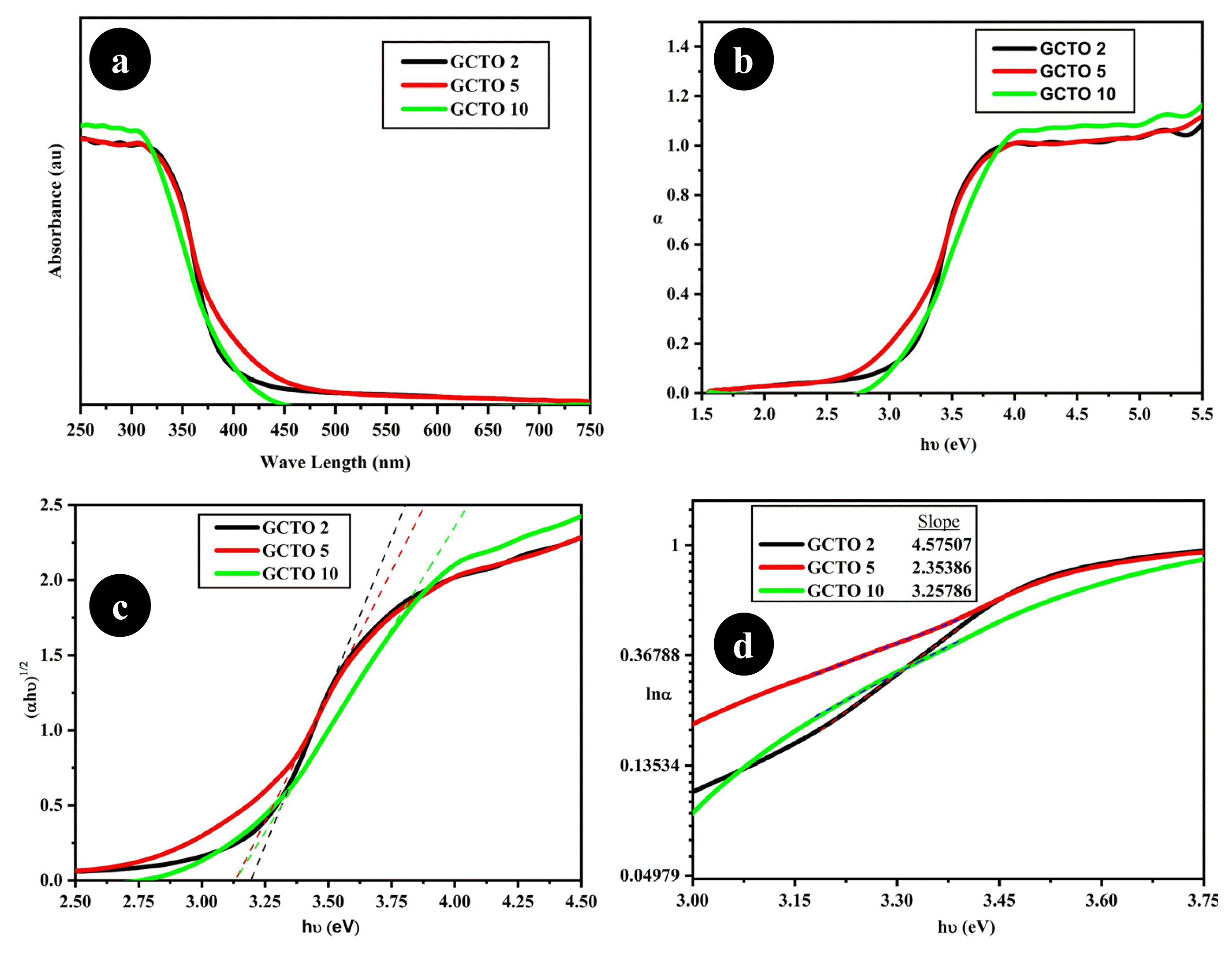
4.3. UV-Visible DRS Studies (Optical Property Study)UV-Visible spectra of GCTO 2, GCTO 5 and GCTO 10 are shown in the Fig. 3(a). The coefficient of absorption (α) and optical band gap (Eg) are related by the equation [53] given below where ‘β’ is called as the proportionality constant and corresponding band gap calculation is done by Tauc’s method.
Similarly, we can calculate static structural disorder [54] through Urbach energy (Eu) calculation. It signifies the induced disorder for high quality semiconductor with crystallinity and becomes larger for highly doped materials due to the contribution from both structural and thermal disorders.
More predominantly it can be expressed by the relation outlined below in equation.
All the values for Eg and Eu for respective samples are given in the Table 2. By comparing the inference retrieved from XRD with UV-Visible DRS data it is evident that the proper doping for GCTO 5 is predominant over GCTO 2 and GCTO 10. The reciprocal of the slope of the liner portion in the figure gives the value of Eu shown in the Fig. 3.
The Table 2 also reveals that the band gap of GCTO 5 in comparison to GCTO 2 is lower due to the doping of Ce4+ into the crystal lattice of TiO2 where excess doping can cause lattice deformation making the crystal less effective towards photocatalytic ability.
4.4. FESEM StudyTo evaluate the morphological characteristics, FESEM study has been carried out for the as prepared composite GCTO 5. Fig. 4 clearly depicts the nano-flakes size of the composite where metal oxide is well stacked into the r-GO sheets. To get better confirmation regarding nanocomposite, elemental analysis is performed through EDAX technique shown in the Fig. 4(c). Atomic percentage of C, O, Ti, and Ce as measured to be 4.56%, 22.32%, 69.54% and 3.59%. Further no other impurity has been noticed as confirmed from the non-availability of any other characteristic peaks in the EDAX spectrum. Hence the method of synthesising material and composite formation has been done at par optimum level to be used for further study in different application.
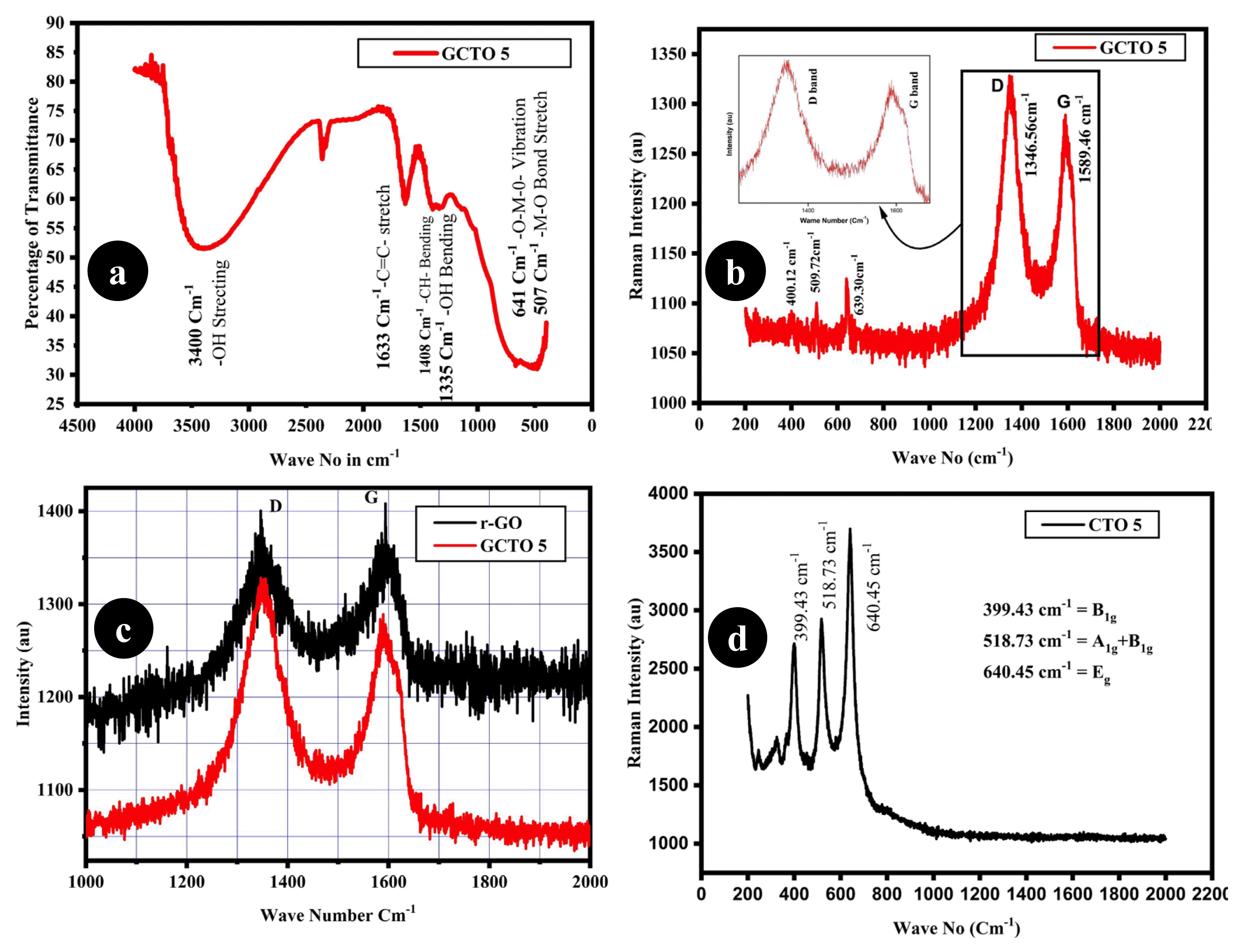
4.5. FTIR and RAMAN AnalysisThe mixed oxide nanocomposites GCTO 5 is subjected to FTIR analysis at 4000 cm−1 to 400 cm−1, and the spectra is being reported in the Fig. 5(a). The broad band spectra at 3400 cm−1 are due to the −OH stretching modes and the nature of widening may be due to the surface attached hydroxyl group. The corresponding peak at 1335 cm−1 is attributed to the in-plane bending vibration of −OH bond. Intense signal in the range of frequency 400 cm−1 to 700 cm−1 clearly signifies the presence of metal oxide and this is due to −M-O lattice vibration and may be due to the −M-O stretching and bending modes. This can also be mapped and well agreement with the RAMAN analysis that the characteristic −MO frequencies are being observed towards slightly lower frequency range indicating the formation of the composite. The signals at 1633 cm−1 and 1408 cm−1 attribute to −C=C-stretching and −CH-bending modes of the r-GO graphitic framework. The signals at 2400 cm−1 may be due to the atmospheric trapping of CO2.
RAMAN scattering technique is a useful tool to get the information about local structures within the composites. To make it clear and to confirm the composite formation with in GCTO 5 between metal oxide and r-GO, this analytical technique has been adopted. In the spectra of Fig. 5(b) the two prominent and highly intense peaks are found at 1346.56 cm−1 and 1589.46 cm−1 can be attributed to the D and G bands respectively. The corresponding D and G band peaks are due to in-plane vibration of k-point phonon for A1g symmetry and first order scattering of E2g phonon due to phase stretching of sp2 carbon atom [55, 56]. The RAMAN active modes in Fig. 5(b) located at 400.12 cm−1, 509.72 cm−1 and 633.30 cm−1 are due to the internal vibration of the mixed metal oxides and responsible for B1g, A1g+B1g and Eg symmetry of anatase having the structural order and disorderness. By comparing the Fig. 5(b) and (c) with Fig. 5(d) and Id/Ig ratio taking into account it can be summarised keeping in view that, the GCTO 5 as synthesised is properly reduced from GO to r-GO along with the composite is formed with metal oxide which is being confirmed by the corresponding RAMAN active modes. A detail pertaining to this is depicted in the Table S2.
4.6. Thermal AnalysisTGA-DTA analysis is carried out to examine the thermal stability of the samples in an inert atmosphere shown in the Fig. 6. It is clearly visualised from the TGA curves for all the sample that the major weight loss is in between 95°C–110°C indicating the loss of entrapped water molecule from the surface of the nanoparticles. No more significant weight loss has been reported in all the sample where maximum 8% for GCTO 2, 8.5% for GCTO 5 and 8.7% for GCTO 10 in total loss is found after the temperature 800°C. It may be due to the increase percentage of cerium loading gradually. A very lower percentage of degradation can be seen between 350°C–500°C indicating the loss of organic functional group like −OH, -C=C-, >C=O and some pyrolytic cleavage of r-GOs. The TGA lines are almost smooth beyond 500°C indicating the electronics application of the sample at higher temperature which is beyond the scope of this paper.
4.7. DLS StudyThis is a supportive technique used for the analysis of hydrodynamic diameter (dnm) of the sample to check the state of agglomeration in due course of its activity during the dispersion in the liquid. All the three samples GCTO 2, GCTO 5 and GCTO 10 are dispersed in DIW and are analysed accordingly. The characteristic peak intensities are given in the Fig. S2. The suspension due to GCTO 5 is accompanied with good hydrodynamic size distribution of 85 nm (dnm) with polydispersity index 0.456 (pdi<0.7) in comparison to other two sample GCTO 2 and GCTO 10.
5. Photocatalytic Activity StudyThe catalytic performance of mixed ceria-titania nanocomposites pertaining to GCTO 2, GCTO 5 and GCTO 10 being investigated by using methylene blue (MB) as a model pollutant under ambient condition in the scale 100 ml of 5ppm MB and 20 mg of the catalyst loading. However, it has been shown that the concentration of MB adsorbed is found to decrease with increasing the concentration CeO2/amount of Ce [57, 58]. Because on increasing the amount of CeO2, the thickness of CeO2 layer becomes higher and the electron and holes created on the CeO2 at the surface gets recombined before the electrons travel and reach the CeO2-TiO2 interface. In our study we have realised that the photocatalytic efficiency is increased with CeO2 loading up to 5% to that of TiO2 in the nanocomposite GCTO 5 and the graphene has also played a key role due to its unique property of high electrical conductivity trapped the excited electron and retarded the electron hole pair recombination and thereby increasing the photocatalytic activity whereas moving towards 10% loading of CeO2 with that of TiO2 again come under less effective towards photocatalytic degradation due to its structural stress. Comparative photocatalytic MB decomposition of the synthesised material is carried out and presented afterwards. All the characterised graphs and pictures are presented in Fig. S3.
The catalytic performance of GCTO 5 would be higher by giving the degradation efficiency of 78.8% at pH=10 for 75 minutes based upon the absorbance noticed for MB at 664nm. The proposed mechanistic path of photocatalytic degradation of MB is as follows:
The semiconducting material GCTO 5 can absorb visible light irradiation and photo generated electron produced being transferred to the conduction band of the corresponding metal oxide of the composite material and due to this inherent property in presence of photon of light, the free electrons in CB and in the dispersed r-GO sheet react in a fashion so that O2 will produce superoxide radical O2.− and the holes h+ being reacted with OH− to generate hydroxyl radicalOH. collectively called as ROS responsible for the degradation of MB [59–63].
5.1. Effect of pHBy moving from pH-3 to pH-10, the catalytic performance of the sample gradually increases by giving higher percentage of degradation after 75 minutes of time. The facts of the finding can be attributed by this inference that, at more acidic condition or less pH the superoxide radical being formed during the photocatalysis encountered by the H+ and due to lower percentage of O2.− yield the potentiality of the catalyst is low at lower pH and higher at pH-10. This result again comes with lower degradation of MB by going beyond the pH-10. At higher concentration of OH−, the photo induced h+ being reacted with OH− to produce OH. where the generated OH. has lower significance in degrading the MB.
5.2. Effect of Competitive Inorganic AnionsThe improvement of photocatalytic performance for GCTO 5 in case of HCO3− with MB in comparison to the degradation of MB without HCO3− is higher may be due to the unique scavenging property of HCO3− for OH. Producing CO3− insisting upon increasing pH from 6 to 7.5. In the counterpart other three anions with GCTO 5 may get reacted with h+ and OH. resulting in the decrease in photocatalytic efficiency.
5.3. LC-MS StudyThe photocatalytic degradation of methylene blue is analysed through LC-MS. The parent peak of MB (with m/z value = 283.56) is obtained. Here the presence of parent peak suggests that there is no complete fragmentation of methylene blue. It is clear from the Fig. S6 that formation of iminium ion is accompanied with demethylation, ring hydroxylation and oxidation of sulphur. The positive mode with m/z corresponds to the values 272, 279, 281, 320, 321, 322 [64]. The respective (M+ = 270) value corresponds to N-demethylation of MB dye due to aerial oxidation to produce Azure B. Within a span of 75 minutes, there is complete degradation of methylene blue with the intermediate’s product formed with m/z values are 255.63, 241.57 and 227.57 g/mol leading to the formulae of azure A, azure C and thionin as shown in Fig. S4 and Fig. S5. Based upon the literature, the photocatalytic degradation of MB may be due to two pathways i.e., chromophore group or the auxochrome group [65–69] which is confirmed by mass spectroscopy. We also expect that the MB degradation has gone through these two pathways with similar m/z values of intermediate products as the peaks associated with m/z 180.75 and 159.76 correspond to the degradation of chromophores [70–73].
5.4. Kinetics Study and Langmuir Adsorption ModelIt is well explained and established that the kinetics of the photocatalytic degradation of MB obeys first order kinetics model [29] and follows the equation mentioned below.
Here kapp is the apparent first order reaction constant, C0 is the initial concentration of the MB solution before photocatalysis, and Ct is the concentration of the MB solution at various time ‘t’. It is clear from the Fig. S7 that the degradation with GCTO 5 would have been superior to GCTO 2 and GCTO 10 as kapp is the measure of the reaction by the formation of more kinetically favourable product. Hence, in this experimental analysis photocatalytic activity of the nanocomposite material GCTO 5 is further studied with varying concentration of MB solution after 75 minutes at pH=10 and corresponding degradation and absorbance intensities are given in Fig. S1(a) with a calibration.
Langmuir adsorption model signifies the qualitative idea about the adsorption capacity of the adsorbent and distribution of adsorbate between solid and liquid phase at equilibrium. The experimental data being found and analysed through this model and given in the Fig. S8 which is well fit with R2=0.993 in accordance with the equation
‘qe’ is the equilibrium concentration, ‘Co’ is the initial concentration of the dye solution, ‘Ce’ is the concentration of dye solution at time 75 minute. ‘V’ is the volume of the solution being taken as 0.1 L; ‘w’ is the weight of the adsorbent where we have taken it as 0.020 g per 0.1 L of the dye solution. KL is the Langmuir constant which can be evaluated out by the relation KL= 1/Slope × qm and the Langmuir constant for our study is found to be 0.3211 L/g. Simultaneously the maximum adsorption capacity of the adsorbent and qm is calculated from the intercept of the line 1/qm and it is found to be 74.1839 mg/g.
6. ConclusionsIn summary, we have fabricated GCTO 5 having percentage variation of CeO2 to that of TiO2 along with composite formation to r-GO, a photo catalyst via sol-gel method. The photocatalytic degradation of MB has been also demonstrated with kinetics phenomenon and according to adsorption model. The presence of graphene with metal oxide in the composite stabilises the anatase phase by increasing the surface area along with visible light absorption and reduces the electron-hole recombination, which in turn improves the photocatalytic activity of the composite material. LC-MS study is successful by carrying out for the detection of methylene blue dye degradation intermediates.
AcknowledgementThe authors are grateful to acknowledge the experimental facilities at the Sophisticated Analytical Instrument Facility, IIT Bombay and CSIR-CIMAP Lucknow. Authors are specially acknowledging S.E.R.B (DST, New Delhi) for funding vide reference no-SR/FST/CS-01/2017/19, Level-1. Priyabrat Mohapatra acknowledges the financial support from Science and Engineering Research Board (SERB), Department of Science and Technology (DST), Government of India, New Delhi, vide File No. EMR/2016/003370.
References1. Lin J, Luo Z, Liu J, Li P. Photocatalytic degradation of methylene blue in aqueous solution by using ZnO-SnO2 nanocomposites. Mater. Sci. Semicond Process. 2018;87:24–31.
http://dx.doi.org/10.1016/j.mssp.2018.07.003
2. Balu S, Uma K, Pan GT, Yang TCK, Ramaraj SK. Degradation of Methylene blue in the presence of visible light using SiO2@α-Fe2O3 Nanocomposites Deposited on SnS2 Flower. Materials. 2018;11(6)1030–1047.
http://dx.doi.org/10.3390/ma11061030
3. Dariani RS, Esmaeili A, Mortezaali A, Dehghanpour S. Photocatalytic reaction and degradation of methylene blue on TiO2 nano-siezed particles. Optik. 2016;127:7143–7154.
http://dx.doi.org/10.1016/j.ijleo.2016.04.026
4. Banerrjee S, Benjwal P, Singh M, Kar KK. Graphene Oxide (rGO) - metal Oxide (TiO2/Fe3O4) based nanocomposites for the removal of methylene blue. Appl. Surf Sci. 2018;489:560–568.
https://doi.org/10.1016/j.apsusc.2018.01.085
5. Li Y, Du Q, Liu T, et al. Comparative study of methylene blue dye adsorption onto activated carbon, graphene oxide and carbon nano tubes. Chem Eng Res Des. 2013;91:361–368.
https://doi.org/10.1016/j.cherd.2012.07.007
6. Hou C, Hu B, Zhu J. Photocatalytic degradation of Methylene blue over TiO2 pre-treated with varying concentration of NaOH. Catalysts. 2018;8:575–588.
https://doi.org/10.3390/catal8120575
7. Panizza M, Cerisola G. Removal of organic pollutant from industrial wastewater by electro generated Fenton's reagent. Water Res. 2001;35:3987–3992.
https://doi.org/10.1016/s0043-1354(01)00135-x
8. Lopez N, Aguila G, Araya P, Guerrero S. Highly active copper based Ce@TiO2 core-shell catalysts for the selective reduction of nitric oxide with carbon monoxide in the presence of oxygen. Catal Commun. 2018;108:17–21.
http://10.1016/j.catcom.2017.10.011
9. Shen L, Xing Z, Zou J, et al. Black TiO2 nanobelts/g-C3N4 nanosheets laminated heterojunctions with efficient visible light driven photocatalytic performance. Sci Rep. 2017;7:1–11.
https://doi.org/10.1038/srep41978
10. Pu S, Zhu R, Ma H, et al. Facile in-situ design strategy to disperse TiO2 nanoparticles on graphene for the enhanced photocatalytic degradation of Rhodamine 6G. Appl. Catal B. 2017;218:208–219.
http://dx.doi.org/10.1016/j.apcatb.2017.06.039
11. Tavakoli F, Badiei A, Ziarani GM, Tarighi S. Photocatalytic application of TiO2-AgI hybrid for degradation of organic pollutants in water. Int. J. Environ Health Res. 2017;11:2017–2024.
https://doi.org/10.1007/s41742-017-0021-7
12. Dominguez S, Huebra M, Han C, et al. Magnetically recoverable TiO2-WO3 photocatalyst to oxidise Bisphenol-A from model wastewater under simulated solar light. Environ. Sci. Pollut Res. 2017;24(14)12589–12598.
https://doi.org/10.1007/s11356-016-7564-6
13. Tabasideh S, Maleki A, Shahmoradi B, Ghahremani E, Mckay G. Sonophotocatalytic degradation of Diazinon in aqueous solution using Iron-doped TiO2 nanoparticles. Sep. Purif Technol. 2017;189:186–192.
https://doi.org/10.1016/j.seppur.2017.07.065
14. Foura G, Soualah A, Robert D. Effect of W-doping level on TiO2 on the photcatalytic degradation of Diuron. Water Sci Technol. 2016;75(1)20–27.
https://doi.org/10.2166/wst.2016.472
15. Li H, Zhou L, Wang L, Liu Y, Lei J, Zhang J. In situ growth of TiO2 nano crystal on g-C3N4 for enhanced photocatalytic performance. Phys. Chem. Chem Phys. 2015;26:1–9.
https://doi.org/10.1039/C5CP02554K
16. Carja G, Husanu E, Gherasi C, Lovu H. New Applications of Nanomaterials. Appl. Catal B. 2011;107:253–259.
https://doi.org/10.1016/j.apcatb.2011.07.020
17. Mohapatra L, Parida KM. Dramatic activities of vanadate intercalated Bismuth doped LDH for solar light photocatalysis. Phys. Chem. Chem Phys. 2014;16:16985–16996.
https://doi.org/10.1039/C4CP01665C
18. Shao M, Han J, Wei M, Evans GD, Duan X. The Synthesis of Hierarchical Zn-Ti Layerd Double Hydroxide for efficient visible light photocatalysis. J. Chem Eng. 2011;168:519–524.
https://doi.org/10.1016/j.cej.2011.01.016
19. Yang X, Cao C, Erickson L, Hohn K, Maghirang R, Klabunde K. Synthesis of visible light active TiO2 based photocatalysts by carbon and nitrogen doping. J Catal. 2008;260:128–133.
https://doi.org/10.1016/j.jcat.2008.09.016
20. Sahibed-dine A, Bentiss F, Bensitel M. The photocatalytic degradation of methylene blue over TiO2 catalysts supported on hydroxyapatite. J Mater. 2017. 841301–1311.
http://www.jmaterenvironsci.com/Journal/vol8-4.html
21. Cabir B, Yurderi M, Caner N, Agirtas MS, Zahmakiran M, Kaya M. Methylene Blue photocatalytic degradation under visible light irradiation on Copper phthalocyanine-sensitised TiO2 nano powders. Mater. Sci. Eng B. 2017;224:9–17.
https://doi.org/10.1016/j.mseb.2017.06.017
22. Dvininov E, Ignat M, Barvinschi P, Smithers MA, Popovici E. New SnO2/MgAl-LDH composites as photocatalysts for cationic dyes bleaching. J. Hazard Mater. 2010;177:150–158.
https://doi.org/10.1016/j.jhazmat.2009.12.011
23. Chen D, Li Y, Zhang J, Li W, Zhou J, Shao L, Qian G. Efficient removal of dyes by a novel magnetic Fe3O4/ZnCr-LDH adsorbent from heavy metal wastewater. J. Hazard Mater. 2012;243:152–160.
https://doi.org/10.1016/j.jhazmat.2012.10.014
24. Hattab AH, Ahmed AI, Hassan SM, Ibrahim AA. Photocatalytic degradation of Methylene Blue by modified nanoparticles titania catalysts. Int. J ModChem. 2015. 7145–53.
https://modernscientificpress.com/Journals/ViewArticle.aspx?H86Z5Noa2iKDNvH/0wRKWvlgO1skO5l/eMv4cRqDh/lhA4mEXohV2jzpAaYmJwh7
25. Sadjadi MS, Mozaffari M, Enhessari M, Zare K. Effect of NiTiO3 nanoparticles supported by mesoporous MCM-41 on photo-reduction of methylene blue under UV-Vis light irradiation. Superlattices Microstruct. 2010;47:685–694.
https://doi.org/10.1016/j.spmi.2010.02.007
26. El-Hakam SA, Abouel-reash YG, Saafan TA, Abd-el-Hameed AG. Photocatalytic degradation of methylene blue by modified bismuth molybdate catalysts. Middle East J. Appl Sci. 2015. 55132–136.
https://www.curresweb.com/mejas/mejas/2015/MEJAS%20Special%20Oct-Dec%202015/132-136.pdf
27. Pandey M, Prajapti R, Shukla P, et al. Synthesis of novel tetra nuclear Ni-complex incorporated mesoporous silica for improved photoMurarilal catalytic degradation of methylene blue in presence of visible light. Polyhedron. 2022;228:116161–116186.
https://doi.org/10.1016/j.poly.2022.116161
28. Duan Y, Shen Y. Synthesis of Zno-CuO/MCM-48 photocatalyst for the degradation of organic pollutions. Water Sci Technol. 2017;76(1)172–181.
https://doi.org/10.2166/wst.2017.196
29. Acosta-silva YJ, Nava R, Hernandez-Morales V, Macias-Sanchez SA, Gomez-Herrera ML, Pawelec B. Methylene blue photo degradation over titania-decorated SBA-15. Appl Catal., B: Environ. 2011;110:108–117.
https://doi.org/10.1016/j.apcatb.2011.08.032
30. Zhai QZ, Dong Y, Liu H, Wang QS. Adsorption of methylene blue on to nano SBA-15 mesoporous material from aqueous media: kinetics, isotherm and thermodynamic studies. Desalin Water Treat. 2019;158:330–342.
http://dx.doi.org/10.1007/s11814-014-0390-y
31. Chakrabarti S, Dutta BK. Photocatalytic degradation of model textile dyes in wastewater using ZnO as semiconductor catalyst. J. Hazard Mater. 2004;B112:269–278.
https://doi.org/10.1016/j.jhazmat.2004.05.013
32. Ke J, Younis MA, Kong Y, et al. Nanostructued ternary metal tungstate based photocatalysts for environmental purification and solar water splitting. Nano-Micro Lett. 2018;69:1–27.
https://doi.org/10.1007/s40820-018-0222-4
33. Pradhan GK, Parida KM. Fabrication of Iron-cerium mixed oxide: an efficient photocatalyst for dye degradation. Eng. Sci. Technol. Int J. 2010;2:53–65.
https://doi.org/10.4314/ijest.v2i8.63780
34. Zhao Z, Tian J, Sang Y, Cabot A, Liu H. Structure, Synthesis, and Application of TiO2 Nanobelts. Adv Mater. 2015;27(16)2557–2582.
https://doi.org/10.1002/adma.201405589
35. Junpoly P, Phuruangrat A, Plubphon N, Thongtem S, Thongtem T. Photocatalytic degradation of methylene blue by Zn2SnO4-SnO2 system under UV visible radiation. Mater Sci Semicond. 2017;66:56–61.
https://doi.org/10.1016/j.mssp.2017.04.010
36. Li Y, Wang W, Wang F, et al. Enhanced photocatalytic degradation of organic dye via defect-rich TiO2 prepared by dielectric barrier discharge plasma. Nanomaterials. 2019;9(5)720–734.
http://dx.doi.org/10.3390/nano9050720
37. Giovannetti R, Rommozzi E, Zannotti M, Amato C. Recent advances in graphene based TiO2 nanocomposite for photocatalytic degradation of synthetic dyes. Catalysts. 2017;7(10)305–339.
https://doi.org/10.3390/catal7100305
38. Arora AK, Jaswal V, Singh K, Singh R. Application of Metal/Mixed metal oxide as photocatalyst. Orient. J Chem. 2016;32:2035–2042.
http://dx.doi.org/10.13005/ojc/320430
39. Abhilash MR, Akshatha G, Srikantaswamy S. Photocatalytic dye degradation and biological activities of the Fe2O3/Cu2O nanocomposite. RSC Adv. 2019;9:8557–8568.
https://doi.org/10.1039%2Fc8ra09929d
40. Ozer N. Optical Properties and electrochemical characterisation of sol-gel deposited ceria films. Sol. Energy Mater Sol Cells. 2001;68:391–400.
https://doi.org/10.1016/S0927-0248(00)00371-8
41. Silva AMT, Silva CG, Drazic G, Faria JL. Ce-doped TiO2 for photocatalytic degradation of chlorophenol. Catal Today. 2001;68:13–18.
https://doi.org/10.1016/j.cattod.2009.02.022
42. Li D, Zhang D, Yu JC. Thermally stable ordered mesoporous CeO2/TiO2 visible-light photocatalysts. Phys. Chem. Chem Phys. 2009;11:3775–3782.
https://doi.org/10.1039/B819167K
43. Xiao J, Peng T, Li R, Peng Z, Yan C. Preparation: phase transformation and photocatalytic activities of cerium doped mesoporous titania nanoparticles. J Solid State Chem. 2006;179:1161–1170.
https://doi.org/10.1016/j.jssc.2006.01.008
44. Pavasupree S, Suzuki Y, Pivsa-Art S, Yoshikawa S. Preparation and characterization of mesoporous TiO2–CeO2 nanopowders respond to visible wavelength. J Solid State Chem. 2005;178:128–134.
https://doi.org/10.1016/j.jssc.2004.10.028
45. Pavasupree S, Suzuki Y, Pivsa-Art S, Yoshikawa S. Synthesis and characterisation of nanoporous, nanorods, nanowires metal oxide. Sci. Technol. Adv Mater. 2005;6:226–229.
https://doi.org/10.1016/j.stam.2005.02.001
46. Huang T, Wu J, Zhao Z, et al. Synthesis and photocatalytic performance of CuO-CeO2/Graphene oxide. Mater Lett. 2016;185:503–506.
https://doi.org/10.1016/j.matlet.2016.09.057
47. Aman N, Satapathy PK, Misra T, Mahato M, Das NN. Synthesis and photocatalytic activity of mesoporous cerium doped TiO2 as visible light sensitive photocatalyst. Mater. Res Bull. 2012;47:179–183.
https://doi.org/10.1016/j.materresbull.2011.11.049
48. Alberoni C, Martin IB, Molina AI, et al. Ceria doping boosts methylene blue photodegradation in titania nanostructure. Mater. Chem Front. 2021;5:4138–4152.
https://doi.org/10.1039/D1QM00068C
49. Djerdj I, Tonejc AM. Structural Investigations of Nanocrystalline TiO2 samples. J Alloys Compd. 2006;413:159–174.
https://doi.org/10.1016/j.jallcom.2005.02.105
50. Satpathy SK, Panigrahi UK, Panda SK, Biswal R, Luyten W, Mallick P. Structural Optical antimicrobial and ferromagnetic properties of Zn1-xLaxO nanorods synthesized by chemical route. J Alloys Compd. 2021;865:158937–158958.
https://doi.org/10.1016/j.jallcom.2021.158937
51. Shi Z, Yang P, Tao F, Zhou R. New insight into the structure of CeO2-TiO2 mixed oxides and their excellent catalytic performance for 1,2-dichloroethane oxidation. J. Chem Eng. 2016;295:99–108.
https://doi.org/10.1016/j.cej.2016.03.032
52. Zhuang X, Li X, Yang Y, et al. Enhanced Sulfamerazine Removal via Adsorption-Photocatalysis Using Bi2O3-TiO2/PAC Ternary Nanoparticles. Water. 2020;12(8)1–18.
https://doi.org/10.3390/w12082273
53. Hafdallah A, Ynineb F, Aida MS, Attaf N. In doped ZnO thin films. J Alloys Compd. 2011;509:7267–7270.
https://doi.org/10.1016/j.jallcom.2011.04.058
54. Cody G, Tiedje T, Abeles B, Moustaks T, Brooks B, Goldstein Y. Disorder and the optical absorption edge of hydrogenated amorphous silicon. J Phys. 1981;42:C4-301–C4-304.
https://doi.org/10.1103/PhysRevLett.47.1480
55. Sahu SC, Samantara AK, Satpati B, Bhattacharjee S, Jena BK. A facile approach for in situ synthesis and graphene-branched-pt hybrid nanostructures with excellent electrochemical performance. Nanoscale. 2013;5:11265–11274.
https://doi.org/10.1039/C3NR03372D
56. Samantara AK, Mishra DK, Suryawanshi SC, et al. Facile synthesis of Ag nanowire-rGO composites and their promising field emission performance. RSC Adv. 2015;52:01–08.
https://doi.org/10.1039/C5RA00308C
57. Noothongkaew S, Thumthan O, An K. Minimal layer graphene/TiO2 nanotube membranes used for enhancement of UV photodetectors. Mater Lett. 2018;218:274–279.
https://doi.org/10.1016/j.matlet.2018.02.033
58. Wang L, Yang W, Chong H, et al. Efficient ultraviolet photodetectors based on TiO2 nanotube array with tailored structures. RSC Adv. 2015;5:52388–52394.
https://doi.org/10.1039/C5RA05861A
59. Molina AI, Villanova A, Talon A, et al. Au-Decorated Ce-Ti Mixed Oxides for Efficient CO Preferential Photooxidation. ACS Appl. Mater Interfaces. 2020;12(34)38019–38030.
https://doi.org/10.1021/acsami.0c08258
60. Ahmadi N, Nemati A, Bagherzadeh M. Synthesis and properties of Ce-doped TiO2 reduced graphene oxide nanocomposite. J Alloys Compd. 2018;742:968–995.
https://doi.org/10.1016/j.jallcom.2018
61. Marcano DC, Kosynkin DV, Berlin JM, Sinitskii A, Sun Z. Improved synthesis of graphene oxide. ACS Nano. 2010;4:4806–4814.
https://doi.org/10.1021/nn1006368
62. Magesh G, Viswanathan B, Viswanath RP, Varadarajan TK. Photocatalytic behaviour of CeO2-TiO2 system for the degradation of methylene blue. Indian J Chem. 2009. 48A:480–488.
https://nopr.niscpr.res.in/bitstream/123456789/3898/1/IJCA%2048A(4)%20480-488.pdf
63. Touati A, Jlaiel L, Najjar W, Sayadi S. Photocatalytic degradation of sulphur black dyes over Ce-TiO2 under UV irradiation: removal efficiency and identification of degraded species. Euro-Mediterr. j. environ integr. 2019;4:1–16.
http://dx.doi.org/10.1007/s41207-018-0086-5
64. Phuruangrat A, Yayapao O, Thongtem T, Thongtem S. Preparation, Characterization, and photocatalytic properties of Ho doped ZnO nanostructures synthesized by Sonochemical Method. Superlattices Microstruct. 2014;67:118–126.
https://doi.org/10.1016/j.spmi.2013.12.023
65. Mohamend RM, Mkhalid IA, Baeissa ES, Rayyani MA. Photocatalytic Degradation of Methylene Blue by Fe/ZnO/SiO2 Nanoparticles under Visible light. J. Nanotechnol. 2012;1–5.
https://doi.org/10.1155/2012/329082
66. Liu G, Wu T, Zhao J, Hidaka H, Serpone N. Photoassisted degradation of dye pollutants. 8. irreversible degradation of Alizarin Red under visible light radiation in air-equilibrated aqueous TiO2 dispersions. Environ. Sci Technol. 1999;33(12)2081–2087.
https://doi.org/10.1021/es9807643
67. Yang S, Cheng Y, Lou LP, Wu X. Involvement of chloride anion in photocatalytic process. J. Environ. Sci (China). 2005. 175761–765.
https://pubmed.ncbi.nlm.nih.gov/16312998/
68. Kuang Y, Zhang X, Zhou S. Adsorption of Methylene Blue in Water onto Activated Carbon by Surfactant Modification. Water. 2020;12:587–606.
https://doi.org/10.3390/w12020587
69. Bakre PV, Volvoikar PS, Vernekar AA, Tilve SG. Influence of acid chain length on the properties of TiO2 prepared by sol-gel method and LC-MS studies of methylene blue photodegradation. J Colloid Interface Sci. 2016;474:58–67.
https://doi.org/10.1016/j.jcis.2016.04.011
70. Wang X, Li Mexi, Xing X, et al. Mechanism and process of methylene blue degradation by manganese oxides under microwave irradiation. Appl. Catal. B. 2014;160–161:211–216.
https://doi.org/10.1016/j.apcatb.2014.05.009
71. Chauaxi Y, Wenping D, Guanevic C, et al. Highly efficient photocatalytic degradation of methylene blue by P2ABSA modified TiO2 nanocomposite due to the photosensitization synergetic effect of TiO2 and P2ABSA. RSC Adv. 2017;7:23699–23708.
https://doi.org/10.1039/C7RA02423A
72. Andriansiferana C, Mohamed EF, Delmas H. Sequential adsorption-photocatalytic oxidation process for wastewater treatment using a composite material TiO2/activated carbon. Environ. Eng Res. 2015;20(2)181–189.
http://dx.doi.org/10.4491/eer.2014.070
73. Mendoza JA, Lee DH, Kang JH. Photocatalytic removal of NOx using TiO2-coated zeolite. Environ. Eng Res. 2016;21(3)291–296.
http://dx.doi.org/10.4491/eer.2016.016
Fig. 1(a) XRD plots for GCTO 2, GCTO 5 and GCTO 10 with corresponding magnifying image (b) GCTO 2 (c) GCTO 5 (d) GCTO 10. 
Fig. 2(a) Adsorption isotherms plot for GCTO 2, GCTO 5 and GCTO 10 (b–d) BJH pore size distribution curve for GCTO 2, GCTO 5 and GCTO 10 (e) multipoint BET plots for GCTO 2, GCTO 5 and GCTO 10. 
Fig. 3(a) UV-Vis DRS spectra (b) RT UV-Vis spectra for GCTO 2, GCTO 5 and GCTO 10, (c) Tauc’s plot for GCTO 2, GCTO 5 and GCTO 10, (d) variation of lnα vs hυ for GCTO 2, GCTO 5 and GCTO 10. 
Fig. 5(a) IR plot for GCTO 5 (b) Raman plot for GCTO 5 (c) Raman plot GCTO 5 and r-GO (d) Raman plot for CTO 5. 
Table 1Different types pollutants with photocatalyst used for the degradation study
Table 2Surface area, pore size/volume distribution, optical band gap energy and Urbach energy of GCTO 2, GCTO 5, GCTO 10
|
|
|||||||||||||||||||||||||||||||||||||||||||||||||||||||||||||||||||||||||||||||||||||||||||||||||||||||||||||||||||||||||||||