


1. Introduction
Pharmaceuticals, surfactants, disinfection by-products, personal care products, pesticides and plasticizers are among chemical compounds of daily use which upon entering into the environment are considered as emerging contaminants (EC) [1]. These pharmaceutical compounds are potential contaminants for human health as well as the environment due to their extensive utilization and persistence. Antibiotics constitute major proportion of the pharmaceutical compounds for human medicine, aquaculture, animal husbandry, veterinary and agriculture [2]. These antibiotics are thus found in all environmental media due to excessive production, utilization and unsafe disposal. The presence of such antibiotics in environment has led to an increase in antibiotic resistance in microbes which is of great concern nowadays. Release of pathogenic bacteria through wastewater addition into the aquatic environments has increased concerns over the ecological impacts of antibiotics [3]. Bacteria that used to be sensitive to antibiotics have now become resistant to a variety of antibacterial agents, mandating improvements in existing disinfection technologies [4, 5]. Recently researchers are trying to develop nanotechnology based water treatment and management systems in search of more efficient and cost effective treatment options against drug resistant pathogens [6, 7]. Enhanced beneficial attributes of nano-sized particles are owed to their surface to volume ratio, structure and morphological features compared to parent materials [8]. The metallic nanomaterials in this scenario have shown notable antibacterial efficiency because of the huge surface areas [9]. The antibacterial activity of nanoparticles has created new application as novel antimicrobial agents that target cell membrane of the resistant microbe in a different manner [10, 11].
Numerous synthesis routes for nanomaterials have been reported by researchers such as solvothermal, sono-chemical, and reverse micro- emulsion showing excellent antimicrobial activity, however, the solvents and other precursors required for in such synthesis routes are unfortunately highly toxic [12]. In order to develop biodegradable and biocompatible nanoparticles a rapid shift has been observed in nanoparticle synthesis strategies from physico-chemical to biological techniques [13, 14] which involve the use of bio-resources like fungi, bacteria and plants for nanoparticles synthesis [15, 16]. Green synthesis for environmental and medical applications is a recently growing technology. It involves utilization of plant borne chemicals for synthesizing nanomaterials as a robust and environment friendly method without high pressure, energy or temperature requirements [17]. In these methods natural reducing agents replace the hazardous or flammable chemical solvents otherwise used in wet chemical synthesis routes to produce metal nanoparticles. Medicinal plants have been considered for producing large number of natural chemical agents that exhibit antioxidant activities. For instance, Ficus palmata leave extracts have been reported to contain alkaloids, tannins, flavonoids, terpenoids and cardiac glycosides [18]. Similarly, leaves of Ficus carica have been reported to possess reducing as well as capping agents that can reduce harmful Ag+ ions in to Ag0 ions [19]. Nano-dimensional silver [20–27] and zinc oxide [28–32] prepared by green synthesis routes have been reported recently with high antibacterial, antifungal, antioxidant and anticancer activities [27–32].
The present work reports the use of Ficus palmate Forsk leaves extracts for the production of zinc oxide and silver nanoparticles as a green synthesis route to be utilized as a potent anti-bacterial remedy against pathogenic and resistant bacteria.
2. Materials and Methods
2.1. Materials
Silver nitrate (AgNO3) and Zinc acetate dihydrate [Zn (CH3COO)2.2H2O] were used as precursors for silver and zinc oxide, respectively. Sodium hydroxide (2M NAOH) solution in deionized water was used for pH adjustment. Plant extract was stored/ preserved at 4°C. The pH of the samples was determined by 8000 Adwa (Hungary Kft.) pH meter. Centrifugation was performed using Jouan centrifuge BR4i (DJB Labcare Ltd England). Memmert UN 55 (Germany) drying oven was used for drying of the nanomaterials. Nutrient agar plate (after the growth of bacterial colonies) incubated at 37°C (Isotemp Oven, Fisher Scientific, USA). Optical density (OD630) of the bacterial inoculums was measured by microplate reader ELx800 (Cole-Parmer, Canada) at 630 nm.
2.2. Test Organisms
Staphylococcus aureus (ATCC-6538), Bacillus subtilis (ATCC-6633), Klebsiella pneumoniae (ATCC-1705), Pseudomonas aeruginosa (ATCC-15442), Escherichia coli (ATCC-25922), Resistant Pseudomonas aeruginosa (MIC-103), Resistant E. coli (MIC-102) and Resistant Streptococcus haemolyticus (MIC-101) (hereafter represented as SA, BS, KP, PA, EC, RPA, REC and RSH, respectively), were provided by Department of Microbiology, Shah Abdul Latif University, Khairpur, Pakistan.
2.3. Synthesis of Silver and Zinc Oxide Nanoparticles
2.3.1. Preparation of plant extract
Ficus palmata leaves were plucked from the trees growing in the gardens of Quaid-i-Azam University and were immediately brought to the Catalysis for Energy and Environment Laboratory, QAU, Pakistan for further processing. Plant leaves were first rinsed with tap water followed by distilled water washing. Leaves were then placed on newspaper and dried under direct sunlight. Dried leaves were then subjected to grounding to obtain fine powder. 10 gm of fine leaf powder was added to 100 mL water followed by 15 min boiling with constant stirring at 500 rpm using magnetic stirrer. The mixture thus obtained was then allowed to cool for some time, was filtered and stored at 4°C [10].
2.3.2. Nanomaterial synthesis
The zinc oxide and silver nanoparticles (ZnO NP and Ag NP, respectively) were prepared as previously reported with some modifications [9, 17]. Zinc accetate (6 g) was added to 60 mL water, 20 mL extract was first boiled for 10 min at 50ºC and Zn (CH3COO)2.2H2O solution was added drop wise. At that time the color of plant extract started fading and pH of the solution mixture was checked which was found to be slightly acidic. It was raised to 11.5 by adding 2M NaOH solution. A complete color change was observed and cream-colored precipitate started forming which was the indication of formation of zinc hydroxide (Zn(OH)2). After centrifugation at 7,000 rpm for 15 min the precipitates were washed, oven dried at 60°C, subsequently ground and stored.
For Ag NP synthesis 10% (w/v) of leaf extract was added to 6 mM AgNO3 1:4 with constant stirring. Appearance of brown color was the indication of Ag+ being reduced to Ag0 and thus Ag NP synthesis [10]. The mixture was then autoclaved (5 min @ 121°C and 15 psi). Thereafter, the mixture was dispersed in distilled water and centrifuged for purification; dried at 80°C and was stored at room temperature for further use.
2.4. Characterization
The morphology and elemental mapping of nanomaterials were determined by using SEM fitted with EDAX using Hitachi S-4800 microscope operated at 20 kV. Prepared nanoparticles were subjected to XRD analysis for determining the crystalline structure with D8, BRUKER AXS diffractometer using Cu Kα radiation (α = 0.15425 nm).
2.5. Antibacterial Activity
The synthesized nanoparticles were tested against 8 different bacterial strains. For these studies, two broad spectrum antibiotics i.e., cefixime and roxithromycin were used as a control group. All the materials and equipment used for antibacterial screening assay were autoclaved. Overnight fresh bacterial cultures were used for the analysis. A suspension was prepared by diluting bacterial colonies in pre-autoclaved nutrient broth in ratio 1: 9. This inoculum was further used in the experiment. Minimum concentration of samples used for growth inhibition of bacteria was investigated by MIC assay [33]. For this threefold dilution of our samples was done.
Where,
The highest concentration comes out to be;
So, four different concentrations of every sample were tested against all bacterial strains i.e. 100, 33.3, 11.1 and 3.7 μg.mL−1. After 24 h incubation the turbidity levels were checked [34].
2.5.1. Antibacterial kinetics
The optical density (OD630) of all bacterial strains was determined to detect the growth with 2 h interval up to 6 h and then at 20, 22 and finally at 24 h. It was done at 630 nm via microplate absorbance reader ELx800. Duplicate experiments were performed [35].
3. Results and Discussion
3.1. Nanomaterial Characterization
Diffraction patterns of ZnO NP are shown in Fig. 1(a) which shows lattice peaks at 31.77°, 34.40°, 36.22°, 47.61°, 56.58°, 62.73°, 67.93°, 69.08°, corresponding to 100, 002, 101, 102, 110, 103, 112, 201 crystal planes, respectively confirming the hexagonal wurtzite structure (JCPDS. 361451) [36]. These ZnO nanoparticles are prominently grown in (101) direction. It was noticed the synthesized sample is well crystalline and single phase with the absence of amorphous part. The lattice parameter a = b = 0.3222 nm and c = 0.5216 nm are calculated and close to earlier reports. Fig. 1(b) shows the well crystalline diffracted XRD patterns at 2θ of values 32.12°, 38.17°, 44.31°, 46.14° and 64.44° that could be attributed to the 98, 111, 200, 101 and 220 planes, respectively. The XRD peaks indicating that structure of synthesized Ag NPs (JCPDS file no. 89-3722), hence it is clear that Ag NP synthesized from Ficus palmata leaf extract were essentially crystalline. The calculated lattice parameters for these Ag NP are a = b = c 0.4048nm and close to previous reports [14].
The microstructure and morphology of the synthesized nanomaterials was obtained from SEM measurements. Fig. 2(a), (b) and (c) shows SEM images Ag NP. Apart from the characteristic bright appearance compered to ZnO NP, the Ag NP have a spherical appearance with particle size 50 to 67 nm. Fig. 2 (d), (e) and (f) shows that the ZnO NP have flake like structure with average particle diameter of 45–60 nm.
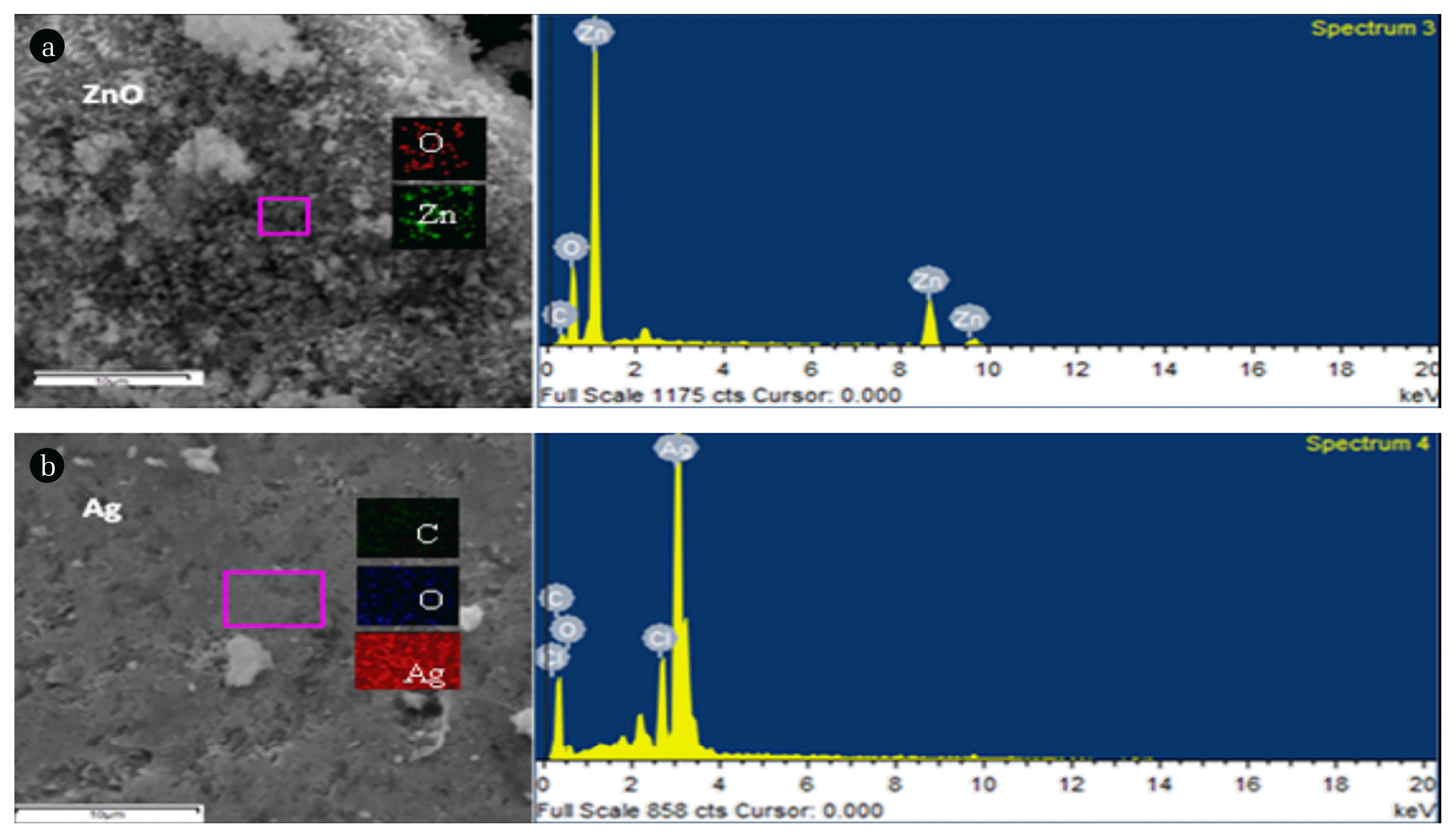
The elemental composition of the synthesized nanomaterials and elemental map (inset) could be elaborated by the representative SEM images coupled with the energy dispersive X-ray spectrum for both nanoparticles (Fig. 3 (a) for ZnO NP and (b) for Ag NP).
3.2. Antibacterial Activity of Nanomaterials
The antimicrobial activities against different pathogens like E. coli, K. pneumoniae, P. aeruginosa, S. aureus, B. subtilis, resistant E. coli, resistant P. aeruginosa and resistant S. haemolyticus were evaluated by determining MIC, kinetics growth curve analysis and growth inhibition percentages. For these studies, two broad spectrum antibiotics i.e., cefixime and roxithromycin were used as a control group.
3.2.1. Minimum inhibitory concentration (MIC) and kinetics growth curve
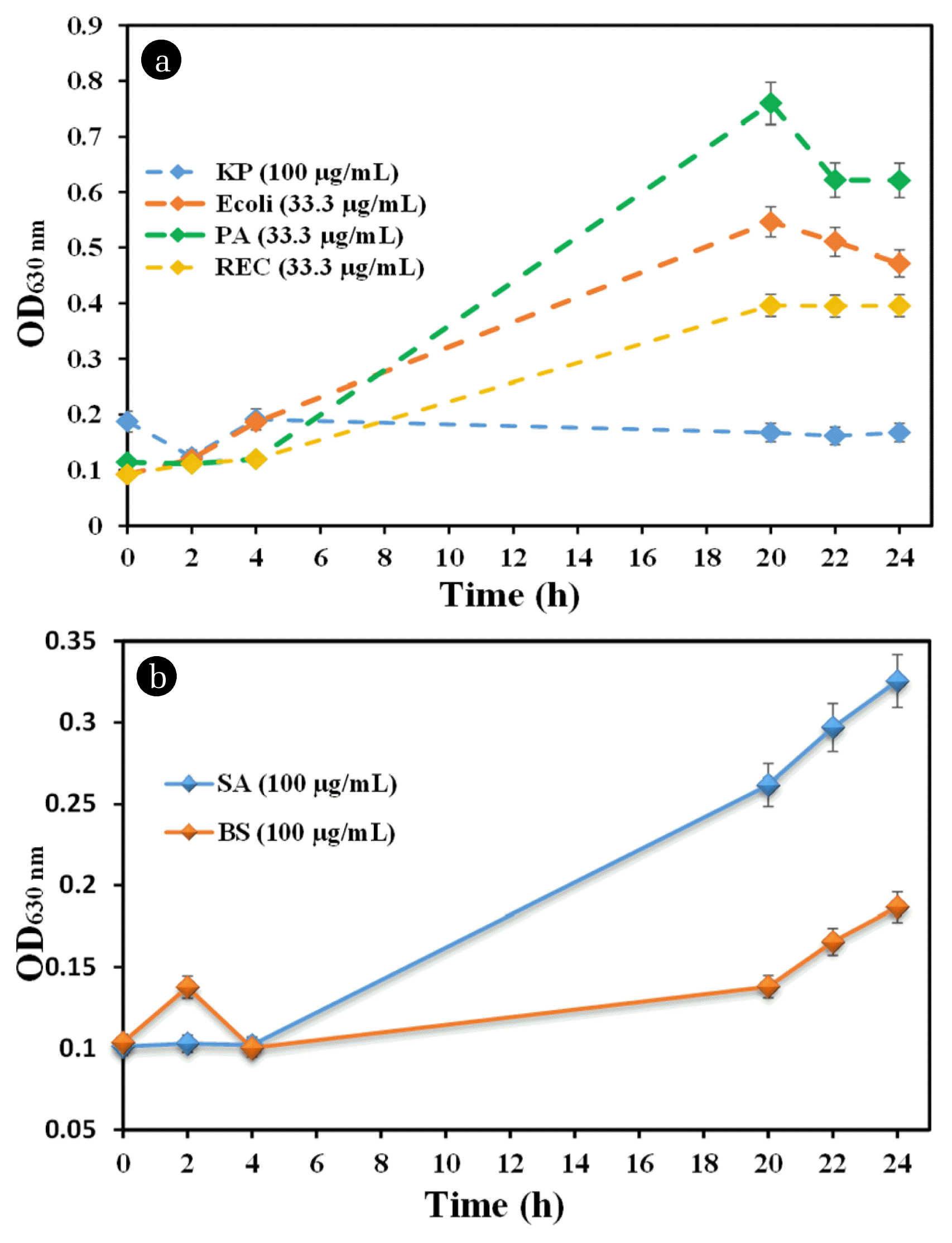
The least amount of dose that is needed to exhibit the bacteriostatic effect is said to be MIC of that sample. Micro-dilution technique was used to determine the MIC of analyzed nanomaterial samples [36]. For Gram positive strains, roxithromycin was tested as control whereas cefixime antibiotic for Gram negative and resistant strains. The activity was checked at four different concentrations which were 100, 33.3, 11.1 and 3.7 μg.mL−1. It was observed that 4 out of 8 bacterial strains were sensitive to the antibiotics tested. The dose of 33.3 μg.mL−1 of cefixime was sufficient for growth inhibition of P. aeruginosa and resistant E. coli. At the same time a larger dose (100 μg.mL−1) of cefixime was required to inhibit K. pneumoniae. Significant bacterial growth was observed at the initial time, however after 20th h a decrease in growth was observed which can be seen in Fig. 4(a). Roxithromycin was found to be ineffective against both bacterial strains (B. subtilis and S. aureus) as shown in Fig. 4(b). All the nanoparticles showed greater activity than the positive control used for this experiment.
3.2.2. Antibacterial silver nanoparticles
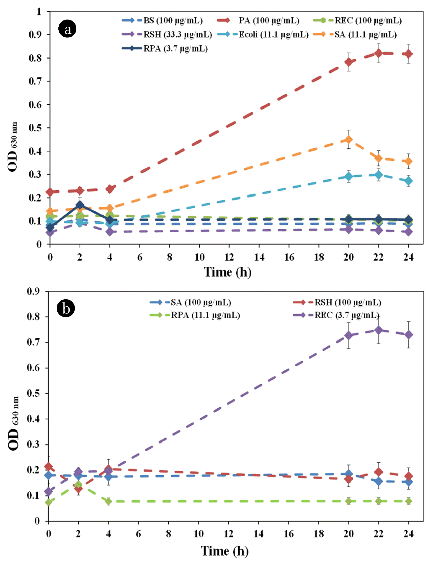
Silver nanoparticles have been reported as alternative antibacterial and antioxidant agents in recent studies [37–39]. Silver nanoparticles (Ag NP) synthesized in this research were also found to be quite effective against bacteria and inhibited growth at different a concentrations. But ineffective against only one bacterial strain K. pneumoniae. For B. subtilis, P. aeruginosa and resistant E. coli the MIC was 100 μg.mL−1. The growth of resistant S. haemolyticus was inhibited by using 33.3 μg.mL−1 Ag NP while 11.1 μg.mL−1 was required for E. coli and S. aureus. Ag NP MIC against resistant P. aeruginosa was the lowest i.e. 3.7 μg.mL−1 as shown in Fig. 5(a). A similar study was conducted by Agnihotri and co-workers in 2014, where significant antibacterial activity was observed by the Ag NP were synthesized via chemical route with MIC reported from 40 to 80 μg.ml−1, respectively [40]. Similarly, Ag NP synthesized from the leaf extract of Alysicarpus monilifer showed greater activity as compared to the antibiotics erythromycin and amoxicillin used for the study [14]. Karunakaran and team synthesized silver and Magnesium nanoparticles from the Hydrangea paniculata flower extract. The synthesized nanoparticles were evaluated for bacterial growth inhibition compared with the antibiotic streptomycin used as a positive control. A greater zone of inhibition was seen for nanoparticles as compared to the positive control [41].
3.2.3. Antibacterial zinc oxide nanoparticles
Fig. 5(b) demonstrates that ZnO NP were found active against 4 strains out of which only one was normal pathogenic strain while remaining three were resistant strains. Its activity was significantly potent against resistant E. coli as 3.7 μg.mL−1 concentration showed inhibition effect, whereas, for Resistant S. haemolyticus and S. aureus 100 μg.mL−1 concentration was needed to inhibit the bacterial growth. An inhibition effect was observed at 11.1 μg.mL−1 concentration against Resistant P. aeruginosa as shown in Fig. 5(b). A similar study conducted in 2015 by Elumalai and Velmurugan showed significantly different results. The ZnO NPs prepared from Azadirachta indica (L.) was active for B. subtilis and E. coli where a dose of 6.25 μg.mL−1 was sufficient for bacterial inhibition. While P. aeruginosa, Proteus mirabilis and E. coli showed growth inhibited at 25 μg.mL−1 [42]. ZnO NP synthesized from Hibiscus rosa-sinensis leaf extract has been reported to show good antibacterial activity against pathogens. It showed better activity against P. aeruginosa whereas least activity was observed against K. pneumoniae [43]. An increase in antibacterial effect of nanoparticle is reported to be related to the miniscule particle size. The antibacterial activity of ZnO NP was also found to be inversely related to its particle size [44]. The MIC of nanoparticles against different bacterial strains is summarized in Table 1.
3.2.4. Percentage growth inhibition
The percentage growth inhibition is defined as:
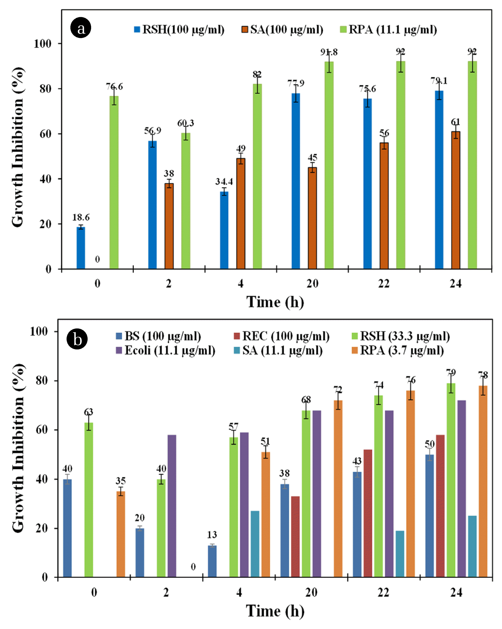
Fig. 6(a) and (b) show the antibacterial activities at specific concentrations of ZnO NP and Ag NP, respectively, against different pathogenic strains. As shown in Fig. 6(a), the ZnO NP were found to be active against resistant S. haemolyticus, resistant P. aeruginosa and K. pneumoniae. With time an increase in growth inhibition was observed. After 20th h the maximum inhibition was observed. ZnO NP showed significant growth inhibition of resistant S. haemolyrticus and resistant P aeruginosa. But even at 100 μg.mL−1 the percent growth inhibition was low against S. aureus. Maximum growth inhibition was observed against resistant S. haemolyticus. For resistant P aeruginosa 11.1 μg.mL−1 concentration worked out quite well.
Silver nanoparticles are well known for the antibacterial effects, therefore, used widely in medical field as well. The Ag NP were potent against bacteria and exhibited antibacterial activity enhancement with time. Fig. 6 shows that growth of most of the strains was inhibited after 20th h of incubation and the percentages were highest at 24th h. The inhibition percentage, however, was not promising for S aureus. As apparent from the Fig. 6(a) and (b), the inhibition percentages of nanoparticles are time dependent. Moghayedi and co-workers reported the mechanism of nanoparticles action over bacteria. It was reported that when treated with nanoparticles bacterial shape initially changes because of the damage caused to their membranes. Afterwards they became inactive and finally die after the leakage of the cytoplasm. Electrostatic attractions between the bacterial cell membrane and the nanoparticles were found to be the reason behind bacterial death [35]. The nanoparticle’s small size and larger surface area can be the reasons behind such low MICs. It is reported that nanoparticles get attach to the cell membranes of bacteria thus disturbing bacterial cellular functions and permeability [44]. Jemal and co-workers reported that positive charge on nanoparticle ions is responsible for higher antimicrobial activity of silver NPs as it gets attached with the negative charge on bacterial membrane through electrostatic forces [9].
4. Conclusion
A rapid, environmentally benign and green route for synthesizing nano-sized zinc oxide and silver is reported using Ficus palmata Forssk leaf extracts. Efficient antibacterial activity of the synthesized nanoparticles was observed against drug resistant and responsive pathogenic bacterial strains. Silver NPs showed great potential against S. aureus, B. subtilis, E. coli, resistant E. coli, resistant P. aeruginosa and resistant S. haemolyticus at MIC concentrations as low as 3.7 μg.mL−1. ZnO NPs also exhibited efficient antibacterial activity for S. aureus and resistant P. aeruginosa even at 11.1 μg.mL−1. The antibacterial activity of synthesized nanomaterials was dose and time dependent. Green synthesized nanoparticles were found to be more effective compared with the antibiotic controls, cefixime and roxithromycin. The reported synthesis route is not only safe, eco-friendly but also provides an alternative route to conventional chemical synthesis of nanomaterials and further can be explored instead of conventional antibiotics.